
PBS, 40% Glycerol, 0.05%BSA, 0.03% Proclin 300
12 months from date of receipt / reconstitution, -20°C as supplied
| 应用 | 稀释度 |
|---|---|
| Dot Blot | 1:1000 |
| WB | 1:1000 |
| IP | 1:50 |
GSK-3 is a serine/threonine protein kinase that phosphorylate either threonine or serine, and this phosphorylation controls a variety of biological activities, such as glycogen metabolism, cell signaling, cellular transport, and others. GSK-3 has also been shown to regulate immune and migratory processes. GSK-3 participates in a number of signaling pathways in the innate immune response, including pro-inflammatory cytokine and interleukin production. GSK-3 is also integrally tied to pathways of cell proliferation and apoptosis. GSK-3 has been shown to phosphorylate Beta-catenin, thus targeting it for degradation. GSK-3 is therefore a part of the canonical Beta-catenin/Wnt pathway, which signals the cell to divide and proliferate. Insulin indirectly inactivates GSK3 via downstream phosphorylation of the specific serine residues Ser21 and Ser9 in GSK-3 isoforms α and β, respectively, via the PI3K/Akt pathway (protein kinase B).
WB result of Phospho-GSK-3β (Ser9) Rabbit mAb
Primary antibody: Phospho-GSK-3β (Ser9) Rabbit mAb at 1/1000 dilution
Lane 1: HeLa whole cell lysate 20 µg
Lane 2: HeLa starve overnight , then treated with Calyculin A (100nM, 30 min) whole cell lysate 20 µg
Secondary antibody: Goat Anti-Rabbit IgG, (H+L), HRP conjugated at 1/10000 dilution
Predicted MW: 47 kDa
Observed MW: 45 kDa
Exposure time: 180 s
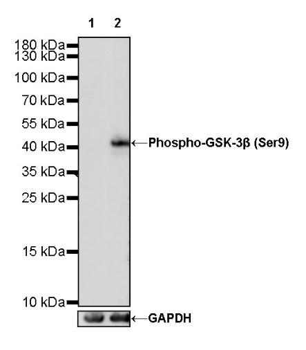
WB result of Phospho-GSK-3β (Ser9) Recombinant Rabbit mAb
Primary antibody: Phospho-GSK-3β (Ser9) Recombinant Rabbit mAb at 1/1000 dilution
Lane 1: Untreated NIH/3T3 whole cell lysate 20 µg
Lane 2: NIH/3T3 starve for 3hr, then treated with Calyculin A (100nM/ml, 30min) whole cell lysate 20 µg
Negative control: Untreated NIH/3T3 whole cell lysate 20 µg
Secondary antibody: Goat Anti-Rabbit IgG, (H+L), HRP conjugated at 1/10000 dilution Predicted MW: 47 kDa
Observed MW: 50kDa
Phospho-GSK-3β (Ser9) Rabbit mAb at 1/50 dilution (1 µg) immunoprecipitating Phospho-GSK-3β (Ser9) in 0.4 mg HeLa starve overnight, then treated with Calyculin A (100nM) for 30 minutes whole cell lysate.
Western blot was performed on the immunoprecipitate using Phospho-GSK-3β (Ser9) Rabbit mAb at 1/1000 dilution.
Secondary antibody (HRP) for IP was used at 1/400 dilution.
Lane 1: HeLa starve overnight, then treated with Calyculin A (100nM) for 30 minutes whole cell lysate 20 µg (Input)
Lane 2: Phospho-GSK-3β (Ser9) Rabbit mAb IP in HeLa starve overnight, then treated with
Calyculin A (100nM) for 30 minutes whole cell lysate
Lane 3: Rabbit monoclonal IgG IP in HeLa starve overnight, then treated with Calyculin A (100nM) for 30 minutes whole cell lysate
Predicted MW: 47 kDa
Observed MW: 47 kDa
Exposure time: 30 s(This blot was developed with high sensitivity substrate)
Dot blot result of Phospho-GSK-3β (Ser9) Rabbit mAb
Lane 1: Phospho-GSK-3β (Ser9) peptide
Lane 2: Phospho-GSK-3β peptide
Primary antibody: Phospho-GSK-3β (Ser9) Rabbit mAb at 1/1000 dilution
Secondary antibody: Goat Anti-Rabbit IgG, (H+L), HRP conjugated at 1/10000 dilution
Exposure time: 18 s
 您现在的位置: