
PBS, 40% Glycerol, 0.05%BSA, 0.03% Proclin 300
12 months from date of receipt / reconstitution, -20 °C as supplied
| 应用 | 稀释度 |
|---|---|
| Dot Blot | 1:1000 |
| WB | 1:1000 |
| IHC-P | 1:500 |
| ICC | 1:500 |
| ChIP | 1:20~1:50 |
H3K9ac is an epigenetic modification to the DNA packaging protein Histone H3. It is a mark that indicates the acetylation at the 9th lysine residue of the histone H3 protein. The H3K9 histone has two jobs. Genes get turned on if this mark is acetylated and silences them if methylated. H3K9ac is an important acetylation and connected with active promoters. H3K9ac and H3K14ac have been shown to be part of the active promoter state. They are also present over bivalent promoters and active enhancers. This is also a mark for liver cancer through a defect in the H3K9ac/H3K9me3 transition.
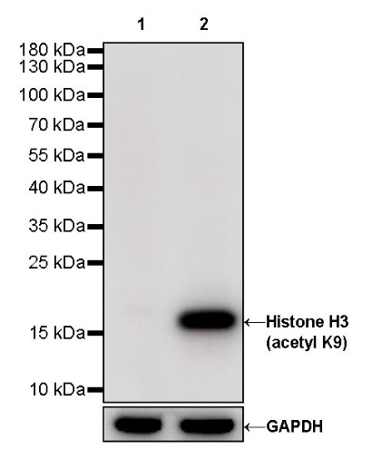
WB result of Histone H3 (acetyl K9) Rabbit pAb
Primary antibody: Histone H3 (acetyl K9) Rabbit pAb at 1/1000 dilution
Lane 1: HeLa whole cell lysate 20 µg
Lane 2: HeLa treated with sodium butyrate (30mM, 4hr) whole cell lysate 20 µg
Secondary antibody: Goat Anti-Rabbit IgG, (H+L), HRP conjugated at 1/10000 dilution
Predicted MW: 15 kDa
Observed MW: 17 kDa
Exposure time: 24 s
WB result of Histone H3 (acetyl K9) Rabbit pAb
Primary antibody: Histone H3 (acetyl K9) Rabbit pAb at 1/1000 dilution
Lane 1: mouse liver lysate 20 µg
Lane 2: mouse spleen lysate 20 µg
Secondary antibody: Goat Anti-Rabbit IgG, (H+L), HRP conjugated at 1/10000 dilution
Predicted MW: 15 kDa
Observed MW: 17 kDa
Exposure time: 180 s
WB result of Histone H3 (acetyl K9) Rabbit pAb
Primary antibody: Histone H3 (acetyl K9) Rabbit pAb at 1/1000 dilution
Lane 1: rat liver lysate 20 µg
Secondary antibody: Goat Anti-Rabbit IgG, (H+L), HRP conjugated at 1/10000 dilution
Predicted MW: 15 kDa
Observed MW: 17 kDa
Exposure time: 180 s
Dot blot result of H3 (acetyl K9) Rabbit pAb
Lane 1: H3 (acetyl K9) peptide
Lane 2: H3 unmodified peptide
Lane 3: H3 (acetyl K27) peptide
Lane 4: H3 (acetyl K4) peptide
Primary antibody: H3 (acetyl K9) Rabbit pAb at 1/1000 dilution
Secondary antibody: Goat Anti-Rabbit IgG, (H+L), HRP conjugated at 1/10000 dilution
Exposure time: 60 s
IHC shows positive staining in paraffin-embedded human stomach. Anti-Histone H3 (acetyl K9) antibody was used at 1/500 dilution, followed by a HRP Polymer for Mouse & Rabbit IgG (ready to use). Counterstained with hematoxylin. Heat mediated antigen retrieval with Tris/EDTA buffer pH9.0 was performed before commencing with IHC staining protocol.
IHC shows positive staining in paraffin-embedded human testis. Anti-Histone H3 (acetyl K9) antibody was used at 1/500 dilution, followed by a HRP Polymer for Mouse & Rabbit IgG (ready to use). Counterstained with hematoxylin. Heat mediated antigen retrieval with Tris/EDTA buffer pH9.0 was performed before commencing with IHC staining protocol.
IHC shows positive staining in paraffin-embedded human breast cancer. Anti-Histone H3 (acetyl K9) antibody was used at 1/500 dilution, followed by a HRP Polymer for Mouse & Rabbit IgG (ready to use). Counterstained with hematoxylin. Heat mediated antigen retrieval with Tris/EDTA buffer pH9.0 was performed before commencing with IHC staining protocol.
IHC shows positive staining in paraffin-embedded human cervical squamous cell carcinoma. Anti-Histone H3 (acetyl K9) antibody was used at 1/500 dilution, followed by a HRP Polymer for Mouse & Rabbit IgG (ready to use). Counterstained with hematoxylin. Heat mediated antigen retrieval with Tris/EDTA buffer pH9.0 was performed before commencing with IHC staining protocol.
IHC shows positive staining in paraffin-embedded mouse colon. Anti-Histone H3 (acetyl K9) antibody was used at 1/500 dilution, followed by a HRP Polymer for Mouse & Rabbit IgG (ready to use). Counterstained with hematoxylin. Heat mediated antigen retrieval with Tris/EDTA buffer pH9.0 was performed before commencing with IHC staining protocol.
IHC shows positive staining in paraffin-embedded rat stomach. Anti-Histone H3 (acetyl K9) antibody was used at 1/500 dilution, followed by a HRP Polymer for Mouse & Rabbit IgG (ready to use). Counterstained with hematoxylin. Heat mediated antigen retrieval with Tris/EDTA buffer pH9.0 was performed before commencing with IHC staining protocol.
ICC shows positive staining in HeLa cells. Anti-Histone H3 (acetyl K9) antibody was used at 1/500 dilution (Green) and incubated overnight at 4°C. Goat polyclonal Antibody to Rabbit IgG - H&L (Alexa Fluor® 488) was used as secondary antibody at 1/1000 dilution. The cells were fixed with 100% ice-cold methanol and permeabilized with 0.1% PBS-Triton X-100. Nuclei were counterstained with DAPI (Blue). Counterstain with tubulin (Red).
Chromatin immunoprecipitation (ChIP) was performed on HeLa cells cross - linked with 1% formaldehyde for 10 min, then chromatin was fragmented by sonication. Parallel reactions used Histone H3 (acetyl K9) Rabbit polyclonal antibody and Rabbit mAb IgG Isotype Control (SDT-R173) at 1:50 for immunoprecipitation. Post - immunoprecipitation, both samples were washed, eluted, and cross - links reversed. Purified DNA was analyzed by qPCR.
qPCR (%input: immunoprecipitated DNA/input DNA) showed the enrichment of RPL30, GAPDH, MYOD1, AFM, SAT-α and SAT-2 in Histone H3 (acetyl K9) Rabbit polyclonal antibody - immunoprecipitated sample.
 您现在的位置: