
12 months from date of receipt / reconstitution, -20 °C as supplied
| 应用 | 稀释度 |
|---|---|
| WB | 1:1000 |
| IHC-P | 1:2000 |
| IF | 1:2000 |
| IP | 1:50 |
AQP-1 is a widely expressed water channel, whose physiological function has been most thoroughly characterized in the kidney. It is found in the basolateral and apical plasma membranes of the proximal tubules, the descending limb of the loop of Henle, and in the descending portion of the vasa recta. Additionally, it is found in red blood cells, vascular endothelium, the gastrointestinal tract, sweat glands, lungs, and the central nervous system. It is a homotetramer with six bilayer spanning domains and N-glycosylation sites, and functions as both a molecular water channel protein and as a non-selective cation channel gated by cyclic guanosine monophosphate (cGMP).
WB result of AQP1 Rabbit mAb
Primary antibody: AQP1 Rabbit mAb at 1/1000 dilution
Lane 1: mouse kidney unboiled RIPA lysate 20 µg
Lane 2: mouse lung unboiled RIPA lysate 20 µg
Secondary antibody: Goat Anti-Rabbit IgG, (H+L), HRP conjugated at 1/10000 dilution
Predicted MW: 28 kDa
Observed MW: 23, 35 kDa
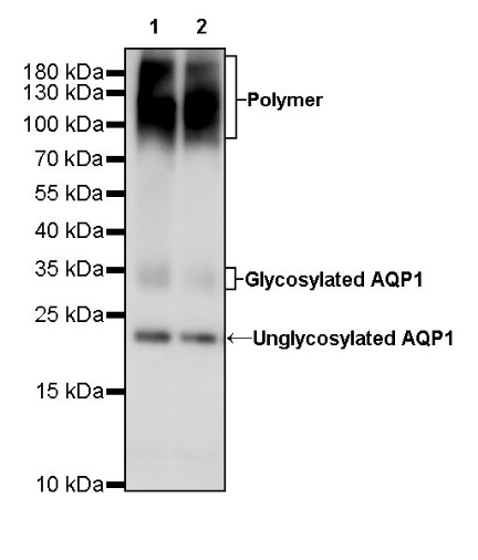
WB result of AQP1 Rabbit mAb
Primary antibody: AQP1 Rabbit mAb at 1/1000 dilution
Lane 1: rat kidney unboiled RIPA lysate 20 µg
Lane 2: rat lung unboiled RIPA lysate 20 µg
Secondary antibody: Goat Anti-Rabbit IgG, (H+L), HRP conjugated at 1/10000 dilution
Predicted MW: 28 kDa
Observed MW: 23, 35 kDa
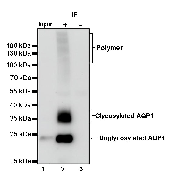
AQP1 Rabbit mAb at 1/50 dilution (1 µg) immunoprecipitating AQP1 in 0.4 mg rat kidney lysate.
Western blot was performed on the immunoprecipitate using AQP1 Rabbit mAb at 1/1000 dilution.
Secondary antibody (HRP) for IP was used at 1/400 dilution.
Lane 1: rat kidney lysate 20 µg (Input)
Lane 2: AQP1 Rabbit mAb IP in rat kidney lysate
Lane 3: Rabbit monoclonal IgG IP in rat kidney lysate
Predicted MW: 28 kDa
Observed MW: 23, 35 kDa
IHC shows positive staining in paraffin-embedded human kidney. Anti-AQP1 antibody was used at 1/2000 dilution, followed by a HRP Polymer for Mouse & Rabbit IgG (ready to use). Counterstained with hematoxylin. Heat mediated antigen retrieval with Tris/EDTA buffer pH9.0 was performed before commencing with IHC staining protocol.
IHC shows positive staining in paraffin-embedded human placenta. Anti-AQP1 antibody was used at 1/2000 dilution, followed by a HRP Polymer for Mouse & Rabbit IgG (ready to use). Counterstained with hematoxylin. Heat mediated antigen retrieval with Tris/EDTA buffer pH9.0 was performed before commencing with IHC staining protocol.
IHC shows positive staining in paraffin-embedded human lung squamous cell carcinoma. Anti-AQP1 antibody was used at 1/2000 dilution, followed by a HRP Polymer for Mouse & Rabbit IgG (ready to use). Counterstained with hematoxylin. Heat mediated antigen retrieval with Tris/EDTA buffer pH9.0 was performed before commencing with IHC staining protocol.
IHC shows positive staining in paraffin-embedded mouse kidney. Anti-AQP1 antibody was used at 1/2000 dilution, followed by a HRP Polymer for Mouse & Rabbit IgG (ready to use). Counterstained with hematoxylin. Heat mediated antigen retrieval with Tris/EDTA buffer pH9.0 was performed before commencing with IHC staining protocol.
IHC shows positive staining in paraffin-embedded rat kidney. Anti-AQP1 antibody was used at 1/2000 dilution, followed by a HRP Polymer for Mouse & Rabbit IgG (ready to use). Counterstained with hematoxylin. Heat mediated antigen retrieval with Tris/EDTA buffer pH9.0 was performed before commencing with IHC staining protocol.
IF shows positive staining in paraffin-embedded human kidney. Anti-AQP1 antibody was used at 1/2000 dilution (Green) and incubated overnight at 4°C. Goat polyclonal Antibody to Rabbit IgG - H&L (Alexa Fluor® 488) was used as secondary antibody at 1/1000 dilution. Counterstained with DAPI (Blue). Heat mediated antigen retrieval with EDTA buffer pH9.0 was performed before commencing with IF staining protocol.
 您现在的位置: