
2 to 8 °C for 2 weeks under sterile conditions;
-20 °C for 3 months under sterile conditions;
-80 °C for 24 months under sterile conditions.
Please avoid repeated freeze-thaw cycles.
| 应用 | 稀释度 |
|---|---|
| FCM | 1:500 |
This antibody recognizes the same epitope as clone 17A2.
The CD3 protein is a T-cell marker, a complex of four structurally distinct membrane glycoprotein isoforms, 20-50 kDa, comprising extracellular, transmembrane and intracellular domains. CD3 is associated with an heterodimer creating the T-cell receptor (TCR). The CD3 complex is responsible for mediating signal transduction to the internal environment upon antigenic recognition by TCR, causing T-cell proliferation and release of cytokines.
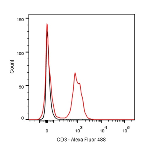
Flow cytometric analysis of Mouse blood cells labelling CD3 antibody at 1/500 (0.1 μg) dilution/ (Red) compared with a Rat monoclonal IgG (Black) isotype control. Goat Anti-Rat IgG Alexa Fluor® 488 was used as the secondary antibody.
Flow cytometric analysis of Mouse splenocytes labelling CD3 antibody at 1/500 (0.1 μg) dilution/ (Red) compared with a Rat monoclonal IgG (Black) isotype control. Goat Anti-Rat IgG Alexa Fluor® 488 was used as the secondary antibody.
 您现在的位置: