
PBS, 40% Glycerol, 0.05%BSA, 0.03% Proclin 300
12 months from date of receipt / reconstitution, -20 °C as supplied
| 应用 | 稀释度 | 推荐种属 |
|---|---|---|
| WB | 1:5000 | Hu |
| IHC-P | 1:500 | Hu |
All mammalian cells express 3 closely related Ras proteins, termed H-Ras, K-Ras, and N-Ras, that promote oncogenesis when they are mutationally activated at codon 12, 13, or 61. N-Ras mutations were more strongly associated with hematopoietic tumors.
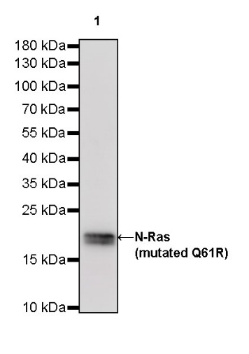
WB result of N-Ras (mutated Q61R) Rabbit mAb
Primary antibody: N-Ras (mutated Q61R) Rabbit mAb at 1/5000 dilution
Lane 1: SK-MEL-2 whole cell lysate 20 µg
Secondary antibody: Goat Anti-Rabbit IgG, (H+L), HRP conjugated at 1/10000 dilution
Predicted MW: 21 kDa
Observed MW: 20 kDa
WB result of N-Ras (mutated Q61R) Rabbit mAb
Primary antibody: N-Ras (mutated Q61R) Rabbit mAb at 1/5000 dilution
Lane 1: 293T transfected with empty vector whole cell lysate 20 µg
Lane 2: 293T transfected with N-Ras-Myc-His fusion protein whole cell lysate 20 µg
Lane 3: 293T transfected with N-Ras (mutated Q61R)-Myc-His fusion protein whole cell lysate 20 µg
Secondary antibody: Goat Anti-Rabbit IgG, (H+L), HRP conjugated at 1/10000 dilution
Predicted MW: 25 kDa
Observed MW: 25 kDa
IHC shows positive staining in paraffin-embedded human melanoma. Anti-N-Ras (mutated Q61R) antibody was used at 1/500 dilution, followed by a HRP Polymer for Mouse & Rabbit IgG (ready to use). Counterstained with hematoxylin. Heat mediated antigen retrieval with Tris/EDTA buffer pH9.0 was performed before commencing with IHC staining protocol.
 您现在的位置: