
12 months from date of receipt / reconstitution, -20°C as supplied
| 应用 | 稀释度 |
|---|---|
| WB | 1:1000 |
| IP | 1:50 |
| IHC-P | 1:500 |
CD82 is a membrane glycoprotein that is a member of the tetraspanin/transmembrane 4 superfamily. Expression of this protein has been shown to be downregulated in tumor progression of human cancers and can be activated by p53 through a consensus binding sequence in the promoter. Its expression and that of p53 are strongly correlated, and the loss of expression of these two proteins is associated with poor survival for prostate cancer patients.
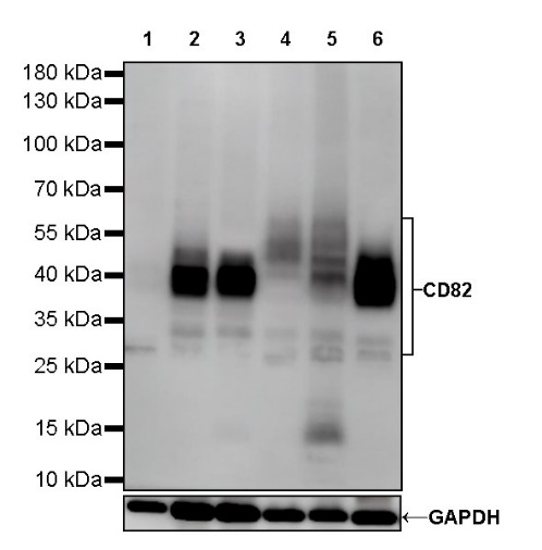
WB result of CD82 Rabbit mAb
Primary antibody: CD82 Rabbit mAb at 1/1000 dilution
Lane 1: SK-OV-3 whole cell lysate 20 µg
Lane 2: Raji whole cell lysate 20 µg
Lane 3: U-87 MG whole cell lysate 20 µg
Lane 4: SW480 whole cell lysate 20 µg
Lane 5: BxPC-3 whole cell lysate 20 µg
Lane 6: Jurkat whole cell lysate 20 µg
Low expression control: SK-OV-3 whole cell lysate
Secondary antibody: Goat Anti-Rabbit IgG, (H+L), HRP conjugated at 1/10000 dilution
Predicted MW: 30 kDa
Observed MW: 30~60 kDa
WB result of CD82 Rabbit mAb
Primary antibody: CD82 Rabbit mAb at 1/1000 dilution
Lane 1: mouse liver lysate 20 µg
Secondary antibody: Goat Anti-Rabbit IgG, (H+L), HRP conjugated at 1/10000 dilution
Predicted MW: 30 kDa
Observed MW: 38~50 kDa
WB result of CD82 Rabbit mAb
Primary antibody: CD82 Rabbit mAb at 1/1000 dilution
Lane 1: rat liver lysate 20 µg
Secondary antibody: Goat Anti-Rabbit IgG, (H+L), HRP conjugated at 1/10000 dilution
Predicted MW: 30 kDa
Observed MW: 40~50 kDa
CD82 Rabbit mAb at 1/50 dilution (1 µg) immunoprecipitating CD82 in 0.4 mg U-87 MG whole cell lysate.
Western blot was performed on the immunoprecipitate using CD82 Rabbit mAb at 1/1000 dilution.
Secondary antibody (HRP) for IP was used at 1/400 dilution.
Lane 1: U-87 MG whole cell lysate 10 µg (Input)
Lane 2: CD82 Rabbit mAb IP in U-87 MG whole cell lysate
Lane 3: Rabbit monoclonal IgG IP in U-87 MG whole cell lysate
Predicted MW: 30 kDa
Observed MW: 30~60 kDa
IHC shows positive staining in paraffin-embedded human tonsil. Anti-CD82 antibody was used at 1/500 dilution, followed by a HRP Polymer for Mouse & Rabbit IgG (ready to use). Counterstained with hematoxylin. Heat mediated antigen retrieval with Tris/EDTA buffer pH9.0 was performed before commencing with IHC staining protocol.
IHC shows positive staining in paraffin-embedded human stomach. Anti-CD82 antibody was used at 1/500 dilution, followed by a HRP Polymer for Mouse & Rabbit IgG (ready to use). Counterstained with hematoxylin. Heat mediated antigen retrieval with Tris/EDTA buffer pH9.0 was performed before commencing with IHC staining protocol.
IHC shows positive staining in paraffin-embedded human cervical squamous cell carcinoma. Anti-CD82 antibody was used at 1/500 dilution, followed by a HRP Polymer for Mouse & Rabbit IgG (ready to use). Counterstained with hematoxylin. Heat mediated antigen retrieval with Tris/EDTA buffer pH9.0 was performed before commencing with IHC staining protocol.
IHC shows positive staining in paraffin-embedded human ovarian carcinoma. Anti-CD82 antibody was used at 1/500 dilution, followed by a HRP Polymer for Mouse & Rabbit IgG (ready to use). Counterstained with hematoxylin. Heat mediated antigen retrieval with Tris/EDTA buffer pH9.0 was performed before commencing with IHC staining protocol.
IHC shows positive staining in paraffin-embedded mouse stomach. Anti-CD82 antibody was used at 1/500 dilution, followed by a HRP Polymer for Mouse & Rabbit IgG (ready to use). Counterstained with hematoxylin. Heat mediated antigen retrieval with Tris/EDTA buffer pH9.0 was performed before commencing with IHC staining protocol.
IHC shows positive staining in paraffin-embedded rat colon. Anti-CD82 antibody was used at 1/500 dilution, followed by a HRP Polymer for Mouse & Rabbit IgG (ready to use). Counterstained with hematoxylin. Heat mediated antigen retrieval with Tris/EDTA buffer pH9.0 was performed before commencing with IHC staining protocol.
 您现在的位置: