
PBS, 0.1% BSA, 0.01% Proclin 300
| 应用 | 稀释度 |
|---|---|
| FCM | 5 μl per million cells in 100μl volume |
CD3 (cluster of differentiation 3) is a protein complex and T cell co-receptor that is involved in activating both the cytotoxic T cell (CD8+ naive T cells) and T helper cells (CD4+ naive T cells). It is composed of four distinct chains. In mammals, the complex contains a CD3γ chain, a CD3δ chain, and two CD3ε chains. These chains associate with the T-cell receptor (TCR) and the CD3-zeta (ζ-chain) to generate an activation signal in T lymphocytes. The TCR, CD3-zeta, and the other CD3 molecules together constitute the TCR complex. The CD3–T cell receptor (TCR) complex plays a central role in the T-cell-mediated immunoresponse as it is involved in the recognition of antigens and subsequent signal transduction and activation of immunocompetent T lymphocytes. Because CD3 is required for T cell activation, drugs (often monoclonal antibodies) that target it are being investigated as immunosuppressant therapies (e.g., otelixizumab, teplizumab) for type 1 diabetes and other autoimmune diseases.
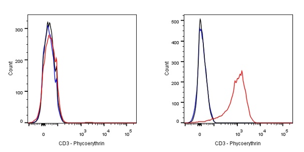
Flow cytometric analysis of CD3 expression on Jurkat. Cells from the Jurkat (Human T cell leukemia T lymphocyte, Right) or Ramos (Human Burkitt's lymphoma B lymphocyte, Left) was stained with Phycoerythrin Mouse IgG2a, κ Isotype Control (Black line histogram) and SDT PE Mouse Anti-Human CD3 antibody (Red line histogram) at 0.1 μg/test, cells without incubation with primary antibody and secondary antibody (Blue line histogram) was used as unlabelled control. Flow cytometry and data analysis were performed using BD FACSymphony™ A1 and FlowJo™ software.
 您现在的位置: