
12 months from date of receipt / reconstitution, -20 °C as supplied
| 应用 | 稀释度 |
|---|---|
| WB | 1:1000 |
| IHC-P | 1:500 |
CD29 is an integrin component that mediates adhesion and involves in homing to sites of inflammation. It expresses in fibroblasts, platelets, T cells, monocytes, granulocytes(low), mast cells, endothelial cells and myoepithelium, also other diverse cell types. It does not express in red blood cells and spermatogonia. It is a myoepithelial marker, although established markers (SMA, CD10, p63, S100, maspin, calponin, GFAP, smooth muscle myosin) are more commonly used.
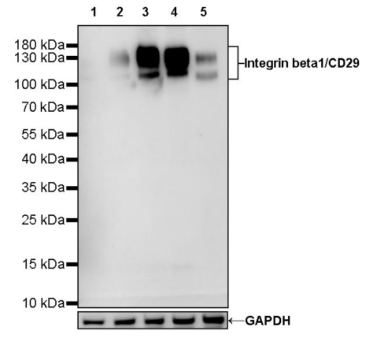
WB result of Integrin beta1/CD29 Rabbit mAb
Primary antibody: Integrin beta1/CD29 Rabbit mAb at 1/1000 dilution
Lane 1: 293T whole cell lysate 20 µg
Lane 2: HeLa whole cell lysate 20 µg
Lane 3: A431 whole cell lysate 20 µg
Lane 4: HT-1080 whole cell lysate 20 µg
Lane 5: MCF7 whole cell lysate 20 µg
Negative control: 293T whole cell lysate
Secondary antibody: Goat Anti-Rabbit IgG, (H+L), HRP conjugated at 1/10000 dilution
Predicted MW: 88 kDa
Observed MW: 110~140 kDa
IHC shows positive staining in paraffin-embedded human skin. Anti-Integrin beta1/CD29antibody was used at 1/500 dilution, followed by a HRP Polymer for Mouse & Rabbit IgG (ready to use). Counterstained with hematoxylin. Heat mediated antigen retrieval with Tris/EDTA buffer pH9.0 was performed before commencing with IHC staining protocol.
IHC shows positive staining in paraffin-embedded human cerebral cortex. Anti-Integrin beta1/CD29antibody was used at 1/500 dilution, followed by a HRP Polymer for Mouse & Rabbit IgG (ready to use). Counterstained with hematoxylin. Heat mediated antigen retrieval with Tris/EDTA buffer pH9.0 was performed before commencing with IHC staining protocol.
IHC shows positive staining in paraffin-embedded human breast. Anti-Integrin beta1/CD29antibody was used at 1/500 dilution, followed by a HRP Polymer for Mouse & Rabbit IgG (ready to use). Counterstained with hematoxylin. Heat mediated antigen retrieval with Tris/EDTA buffer pH9.0 was performed before commencing with IHC staining protocol.
IHC shows positive staining in paraffin-embedded human colon. Anti-Integrin beta1/CD29antibody was used at 1/500 dilution, followed by a HRP Polymer for Mouse & Rabbit IgG (ready to use). Counterstained with hematoxylin. Heat mediated antigen retrieval with Tris/EDTA buffer pH9.0 was performed before commencing with IHC staining protocol.
IHC shows positive staining in paraffin-embedded human kidney. Anti-Integrin beta1/CD29antibody was used at 1/500 dilution, followed by a HRP Polymer for Mouse & Rabbit IgG (ready to use). Counterstained with hematoxylin. Heat mediated antigen retrieval with Tris/EDTA buffer pH9.0 was performed before commencing with IHC staining protocol.
IHC shows positive staining in paraffin-embedded human liver. Anti-Integrin beta1/CD29antibody was used at 1/500 dilution, followed by a HRP Polymer for Mouse & Rabbit IgG (ready to use). Counterstained with hematoxylin. Heat mediated antigen retrieval with Tris/EDTA buffer pH9.0 was performed before commencing with IHC staining protocol.
IHC shows positive staining in paraffin-embedded human placenta. Anti-Integrin beta1/CD29antibody was used at 1/500 dilution, followed by a HRP Polymer for Mouse & Rabbit IgG (ready to use). Counterstained with hematoxylin. Heat mediated antigen retrieval with Tris/EDTA buffer pH9.0 was performed before commencing with IHC staining protocol.
IHC shows positive staining in paraffin-embedded human colon cancer. Anti-Integrin beta1/CD29antibody was used at 1/500 dilution, followed by a HRP Polymer for Mouse & Rabbit IgG (ready to use). Counterstained with hematoxylin. Heat mediated antigen retrieval with Tris/EDTA buffer pH9.0 was performed before commencing with IHC staining protocol.
 您现在的位置: