
12 months from date of receipt / reconstitution, -20 °C as supplied
| 应用 | 稀释度 |
|---|---|
| WB | 1:1000 |
| ICC | 1:100 |
| ICFCM | 1:50 |
S-methyl-5'-thioadenosine phosphorylase (MTAP) is an enzyme that plays a major role in polyamine metabolism and is important for the salvage of both adenine and methionine. It is responsible for the first step in this pathway, where it catalyzes the reversible phosphorylation of MTA to adenine and 5-methylthioribose-1-phosphate. This takes place after MTA is generated from S-adenosylmethionine.
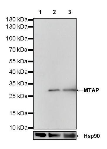
WB result of MTAP Rabbit mAb
Primary antibody: MTAP Rabbit mAb at 1/1000 dilution
Lane 1: A549 whole cell lysate 20 µg
Lane 2: HT-29 whole cell lysate 20 µg
Lane 3: HeLa whole cell lysate 20 µg
Negative control: A549 whole cell lysate
Secondary antibody: Goat Anti-Rabbit IgG, (H+L), HRP conjugated at 1/10000 dilution
Predicted MW: 31 kDa
Observed MW: 31 kDa
WB result of MTAP Rabbit mAb
Primary antibody: MTAP Rabbit mAb at 1/1000 dilution
Lane 1: NIH/3T3 whole cell lysate 20 µg
Lane 2: RAW 264.7 whole cell lysate 20 µg
Secondary antibody: Goat Anti-Rabbit IgG, (H+L), HRP conjugated at 1/10000 dilution
Predicted MW: 31 kDa
Observed MW: 31 kDa
WB result of MTAP Rabbit mAb
Primary antibody: MTAP Rabbit mAb at 1/1000 dilution
Lane 1: C6 whole cell lysate 20 µg
Secondary antibody: Goat Anti-Rabbit IgG, (H+L), HRP conjugated at 1/10000 dilution
Predicted MW: 31 kDa
Observed MW: 31 kDa
Flow cytometric analysis of 4% PFA fixed 90% methanol permeabilized HeLa (Human cervix adenocarcinoma epithelial cell) labelling MTAP antibody at 1/50 dilution (1 μg) / (Red) compared with a Rabbit monoclonal IgG (Black) isotype control and an unlabelled control (cells without incubation with primary antibody and secondary antibody) (Blue). Goat Anti - Rabbit IgG Alexa Fluor® 488 was used as the secondary antibody.
ICC shows positive staining in HeLa cells. Anti-MTAP antibody was used at 1/100 dilution (Green) and incubated overnight at 4°C. Goat polyclonal Antibody to Rabbit IgG - H&L (Alexa Fluor® 488) was used as secondary antibody at 1/1000 dilution. The cells were fixed with 4% PFA and permeabilized with 0.1% PBS-Triton X-100. Nuclei were counterstained with DAPI (Blue). Counterstain with tubulin (red).
 您现在的位置: