
12 months from date of receipt / reconstitution, -20 °C as supplied
| 应用 | 稀释度 |
|---|---|
| WB | 1:1000 |
| IHC-P | 1:500 |
| ICC | 1:500 |
| ICFCM | 1:500 |
Receptor tyrosine-protein kinase erbB-4 is a receptor tyrosine kinase that is a member of the epidermal growth factor receptor family. ERBB4 is a single-pass type I transmembrane protein with multiple furin-like cysteine rich domains, a tyrosine kinase domain, a phosphotidylinositol-3 kinase binding site and a PDZ domain binding motif. The protein binds to and is activated by neuregulins-2, -3 and -4, heparin-binding EGF-like growth factor and betacellulin. Ligand binding induces a variety of cellular responses including mitogenesis and differentiation.
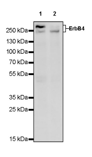
WB result of ErbB4 Rabbit mAb
Primary antibody: ErbB4 Rabbit mAb at 1/1000 dilution
Lane 1: T-47D whole cell lysate 20 µg
Lane 2: MCF7 whole cell lysate 20 µg
Secondary antibody: Goat Anti-Rabbit IgG, (H+L), HRP conjugated at 1/10000 dilution
Predicted MW: 147 kDa
Observed MW: 240, 300 kDa
Flow cytometric analysis of 4% PFA fixed 90% methanol permeabilized T-47D (Human ductal breast epithelial tumor epithelial cell) labelling ErbB4 antibody at 1/500 dilution (0.1 μg) / (Red) compared with a Rabbit monoclonal IgG (Black) isotype control and an unlabelled control (cells without incubation with primary antibody and secondary antibody) (Blue). Goat Anti - Rabbit IgG Alexa Fluor® 488 was used as the secondary antibody.
IHC shows positive staining in paraffin-embedded human breast. Anti-ErbB4 antibody was used at 1/500 dilution, followed by a HRP Polymer for Mouse & Rabbit IgG (ready to use). Counterstained with hematoxylin. Heat mediated antigen retrieval with Tris/EDTA buffer pH9.0 was performed before commencing with IHC staining protocol.
IHC shows positive staining in paraffin-embedded human breast cancer. Anti-ErbB4 antibody was used at 1/500 dilution, followed by a HRP Polymer for Mouse & Rabbit IgG (ready to use). Counterstained with hematoxylin. Heat mediated antigen retrieval with Tris/EDTA buffer pH9.0 was performed before commencing with IHC staining protocol.
IHC shows positive staining in paraffin-embedded human prostatic hyperplasia. Anti-ErbB4 antibody was used at 1/500 dilution, followed by a HRP Polymer for Mouse & Rabbit IgG (ready to use). Counterstained with hematoxylin. Heat mediated antigen retrieval with Tris/EDTA buffer pH9.0 was performed before commencing with IHC staining protocol.
IHC shows positive staining in paraffin-embedded human breast cancer. Anti-ErbB4 antibody was used at 1/500 dilution, followed by a HRP Polymer for Mouse & Rabbit IgG (ready to use). Counterstained with hematoxylin. Heat mediated antigen retrieval with Tris/EDTA buffer pH9.0 was performed before commencing with IHC staining protocol.
ICC shows positive staining in T-47D cells. Anti-ErbB4 antibody was used at 1/500 dilution (Green) and incubated overnight at 4°C. Goat polyclonal Antibody to Rabbit IgG - H&L (Alexa Fluor® 488) was used as secondary antibody at 1/1000 dilution. The cells were fixed with 4% PFA and permeabilized with 0.1% PBS-Triton X-100. Nuclei were counterstained with DAPI (Blue). Counterstain with tubulin (red).
 您现在的位置: