
12 months from date of receipt / reconstitution, -20 °C as supplied
| 应用 | 稀释度 |
|---|---|
| WB | 1:1000 |
| IP | 1:50 |
| IHC-P | 1:100 |
| ICC | 1:500 |
| ICFCM | 1:50 |
PFKFB3 is a gene that encodes the 6-phosphofructo-2-kinase/fructose-2,6-biphosphatase 3 enzyme in humans. It is one of 4 tissue-specific PFKFB isoenzymes identified currently (PFKFB1-4). PFKFB3 converts fructose-6-phosphate to fructose-2,6-bisP (F2,6BP). F2,6BP is a ‘potent’ allosteric activator of 6-phosphofructokinase-1 (PFK-1), stimulating glycolysis. In neurons, glucose is mainly metabolized through the pentose–phosphate pathway (PPP), which is required for NADPH(H+) regeneration and maintenance of neuronal redox status. This neuronal metabolic switch is dictated by the PFKFB3 activity. In neurons, PFKFB3 protein abundance is negligible due to the continuous proteasomal degradation of the enzyme. However, overexcitation of N-methyl-D-aspartate subtype of glutamate receptors (NMDAR), known as excitotoxicity, stabilizes PFKFB3 protein in neurons, resulting in a redirection of glucose flux from PPP to glycolysis, followed by low NADPH(H+) availability for proper GSH regeneration; this ultimately leads to oxidative stress and neuronal death. PFKFB3 is also associated with the Warburg effect because its activity increases the rate of glycolysis. PFKFB3 has been found to be upregulated in numerous cancers, including colon, breast, ovarian, and thyroid.
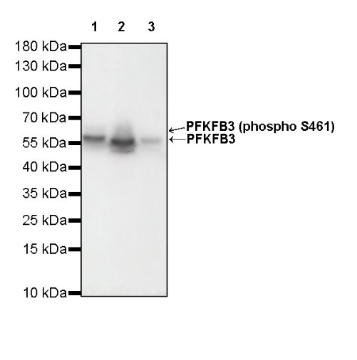
WB result of PFKFB3 Rabbit mAb
Primary antibody: PFKFB3 Rabbit mAb at 1/1000 dilution
Lane 1: A431 whole cell lysate 20 µg
Lane 2: HCT116 whole cell lysate 20 µg
Lane 3: HeLa whole cell lysate 20 µg
Secondary antibody: Goat Anti-Rabbit IgG, (H+L), HRP conjugated at 1/10000 dilution
Predicted MW: 60 kDa
Observed MW: 60 kDa
(This blot was developed with high sensitivity substrate)
WB result of PFKFB3 Rabbit mAb
Primary antibody: PFKFB3 Rabbit mAb at 1/1000 dilution
Lane 1: NIH/3T3 whole cell lysate 20 µg
Secondary antibody: Goat Anti-Rabbit IgG, (H+L), HRP conjugated at 1/10000 dilution
Predicted MW: 60 kDa
Observed MW: 60 kDa
(This blot was developed with high sensitivity substrate)
WB result of PFKFB3 Rabbit mAb
Primary antibody: PFKFB3 Rabbit mAb at 1/1000 dilution
Lane 1: C6 whole cell lysate 20 µg
Secondary antibody: Goat Anti-Rabbit IgG, (H+L), HRP conjugated at 1/10000 dilution
Predicted MW: 60 kDa
Observed MW: 60 kDa
(This blot was developed with high sensitivity substrate)
Flow cytometric analysis of 4% PFA fixed 90% methanol permeabilized A431 (Human epidermoid carcinoma epithelial cell) labelling PFKFB3 antibody at 1/50 dilution (1 μg) / (Red) compared with a Rabbit monoclonal IgG (Black) isotype control and an unlabelled control (cells without incubation with primary antibody and secondary antibody) (Blue). Goat Anti - Rabbit IgG Alexa Fluor® 488 was used as the secondary antibody.
PFKFB3 Rabbit mAb at 1/50 dilution (1 µg) immunoprecipitating PFKFB3 in 0.4 mg A431 whole cell lysate.
Western blot was performed on the immunoprecipitate using PFKFB3 Rabbit mAb at 1/1000 dilution.
Secondary antibody (HRP) for IP was used at 1/400 dilution.
Lane 1: A431 whole cell lysate 10 µg (Input)
Lane 2: PFKFB3 Rabbit mAb IP in A431 whole cell lysate
Lane 3: Rabbit monoclonal IgG IP in A431 whole cell lysate
Predicted MW: 60 kDa
Observed MW: 60 kDa
(This blot was developed with high sensitivity substrate)
IHC shows positive staining in paraffin-embedded human lung squamous cell carcinoma. Anti-PFKFB3 antibody was used at 1/100 dilution, followed by a HRP Polymer for Mouse & Rabbit IgG (ready to use). Counterstained with hematoxylin. Heat mediated antigen retrieval with Tris/EDTA buffer pH9.0 was performed before commencing with IHC staining protocol.
IHC shows positive staining in paraffin-embedded mouse testis. Anti-PFKFB3 antibody was used at 1/100 dilution, followed by a HRP Polymer for Mouse & Rabbit IgG (ready to use). Counterstained with hematoxylin. Heat mediated antigen retrieval with Tris/EDTA buffer pH9.0 was performed before commencing with IHC staining protocol.
IHC shows positive staining in paraffin-embedded rat spleen. Anti-PFKFB3 antibody was used at 1/100 dilution, followed by a HRP Polymer for Mouse & Rabbit IgG (ready to use). Counterstained with hematoxylin. Heat mediated antigen retrieval with Tris/EDTA buffer pH9.0 was performed before commencing with IHC staining protocol.
ICC shows positive staining in A431 cells. Anti-PFKFB3 antibody was used at 1/500 dilution (Green) and incubated overnight at 4°C. Goat polyclonal Antibody to Rabbit IgG - H&L (Alexa Fluor® 488) was used as secondary antibody at 1/1000 dilution. The cells were fixed with 4% PFA and permeabilized with 0.1% PBS-Triton X-100. Nuclei were counterstained with DAPI (Blue). Counterstain with tubulin (Red).
 您现在的位置: