
12 months from date of receipt / reconstitution, -20 °C as supplied
| 应用 | 稀释度 |
|---|---|
| WB | 1:1000 |
| IHC-P | 1:500 |
| ICC | 1:500 |
| ICFCM | 1:500 |
| IP | 1:50 |
HMGA1 is a non-histone chromatin protein involved in many cellular processes, including regulation of inducible gene transcription, DNA replication, heterochromatin organization, integration of retroviruses into chromosomes, and the metastatic progression of cancer cells. Recently it has been shown that HMGA1 proteins, HMGA1a and HMGA1b, can cross-link DNA fibers in vitro and can induce chromatin clustering in vivo suggesting a structural role of HMGA1 proteins in heterochromatin organization.
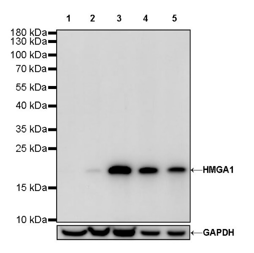
WB result of HMGA1 Rabbit mAb
Primary antibody: HMGA1 Rabbit mAb at 1/1000 dilution
Lane 1: MCF7 whole cell lysate 20 µg
Lane 2: HeLa whole cell lysate 20 µg
Lane 3: NCCIT whole cell lysate 20 µg
Lane 4: SK-OV-3 whole cell lysate 20 µg
Lane 5: MDA-MB-231 whole cell lysate 20 µg
Low expression control: MCF7 whole cell lysate
Secondary antibody: Goat Anti-Rabbit IgG, (H+L), HRP conjugated at 1/10000 dilution
Predicted MW: 12 kDa
Observed MW: 17 kDa
Flow cytometric analysis of 4% PFA fixed 90% methanol permeabilized HeLa (Human cervix adenocarcinoma epithelial cell) labelling HMGA1 antibody at 1/500 dilution (0.1 μg) / (Red) compared with a Rabbit monoclonal IgG (Black) isotype control and an unlabelled control (cells without incubation with primary antibody and secondary antibody) (Blue). Goat Anti - Rabbit IgG Alexa Fluor® 488 was used as the secondary antibody.
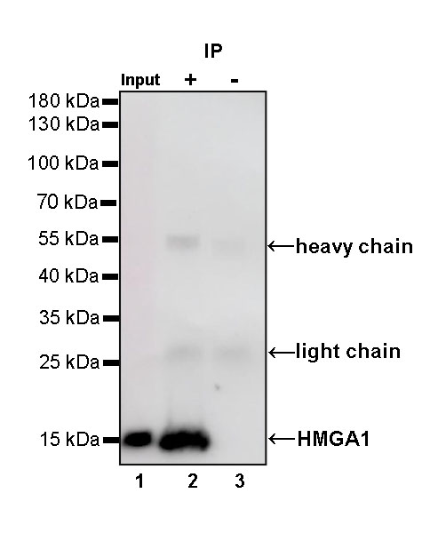
HMGA1 Rabbit mAb at 1/50 dilution (1 µg) immunoprecipitating HMGA1 in 0.4 mg SK-OV-3 whole cell lysate.
Western blot was performed on the immunoprecipitate using HMGA1 Rabbit mAb at 1/1000 dilution.
Secondary antibody (HRP) for IP was used at 1/400 dilution.
Lane 1: SK-OV-3 whole cell lysate 20 µg (Input)
Lane 2: HMGA1 Rabbit mAb IP in SK-OV-3 whole cell lysate
Lane 3: Rabbit monoclonal IgG IP in SK-OV-3 whole cell lysate
Predicted MW: 12 kDa
Observed MW: 16 kDa
IHC shows positive staining in paraffin-embedded human breast cancer. Anti-HMGA1 antibody was used at 1/500 dilution, followed by a HRP Polymer for Mouse & Rabbit IgG (ready to use). Counterstained with hematoxylin. Heat mediated antigen retrieval with Tris/EDTA buffer pH9.0 was performed before commencing with IHC staining protocol.
IHC shows positive staining in paraffin-embedded human colon cancer. Anti-HMGA1 antibody was used at 1/500 dilution, followed by a HRP Polymer for Mouse & Rabbit IgG (ready to use). Counterstained with hematoxylin. Heat mediated antigen retrieval with Tris/EDTA buffer pH9.0 was performed before commencing with IHC staining protocol.
IHC shows positive staining in paraffin-embedded human ovarian cancer. Anti-HMGA1 antibody was used at 1/500 dilution, followed by a HRP Polymer for Mouse & Rabbit IgG (ready to use). Counterstained with hematoxylin. Heat mediated antigen retrieval with Tris/EDTA buffer pH9.0 was performed before commencing with IHC staining protocol.
IHC shows positive staining in paraffin-embedded human pancreatic cancer. Anti-HMGA1 antibody was used at 1/500 dilution, followed by a HRP Polymer for Mouse & Rabbit IgG (ready to use). Counterstained with hematoxylin. Heat mediated antigen retrieval with Tris/EDTA buffer pH9.0 was performed before commencing with IHC staining protocol.
IHC shows positive staining in paraffin-embedded human transitional cell cancer. Anti-HMGA1 antibody was used at 1/500 dilution, followed by a HRP Polymer for Mouse & Rabbit IgG (ready to use). Counterstained with hematoxylin. Heat mediated antigen retrieval with Tris/EDTA buffer pH9.0 was performed before commencing with IHC staining protocol.
ICC shows positive staining in HeLa cells. Anti-HMGA1 antibody was used at 1/500 dilution (Green) and incubated overnight at 4°C. Goat polyclonal Antibody to Rabbit IgG - H&L (Alexa Fluor® 488) was used as secondary antibody at 1/1000 dilution. The cells were fixed with 4% PFA and permeabilized with 0.1% PBS-Triton X-100. Nuclei were counterstained with DAPI (Blue). Counterstain with tubulin (Red).
 您现在的位置: