
12 months from date of receipt / reconstitution, -20 °C as supplied
| 应用 | 稀释度 |
|---|---|
| WB | 1:1000 |
| IP | 1:50 |
| IHC-P | 1:250 |
| ICC | 1:100 |
| ICFCM | 1:50 |
Glutathione S-transferase kappa 1 (GSTK1) is an enzyme that in humans is encoded by the GSTK1 gene which is located on chromosome seven. It belongs to the superfamily of enzymes known as glutathione S-transferase (GST), which are mainly known for cellular detoxification. It has been suggested that, similar to GSTA, GSTK1 may play a role in the buffering of acyl-CoA and xenobiotic-CoA and be involved in their binding activities.
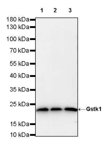
WB result of Gstk1 Rabbit mAb
Primary antibody: Gstk1 Rabbit mAb at 1/1000 dilution
Lane 1: A549 whole cell lysate 20 µg
Lane 2: HeLa whole cell lysate 20 µg
Lane 3: Daudi whole cell lysate 20 µg
Secondary antibody: Goat Anti-Rabbit IgG, (H+L), HRP conjugated at 1/10000 dilution
Predicted MW: 25 kDa
Observed MW: 23 kDa
Flow cytometric analysis of 4% PFA fixed 90% methanol permeabilized A549 (Human lung carcinoma epithelial cell) labelling Gstk1 antibody at 1/50 dilution (1 μg) / (Red) compared with a Rabbit monoclonal IgG (Black) isotype control and an unlabelled control (cells without incubation with primary antibody and secondary antibody) (Blue). Goat Anti - Rabbit IgG Alexa Fluor® 488 was used as the secondary antibody.
Gstk1 Rabbit mAb at 1/50 dilution (1 µg) immunoprecipitating Gstk1 in 0.4 mg A549 whole cell lysate.
Western blot was performed on the immunoprecipitate using Gstk1 Rabbit mAb at 1/1000 dilution.
Secondary antibody (HRP) for IP was used at 1/400 dilution.
Lane 1: A549 whole cell lysate 20 µg (Input)
Lane 2: Gstk1 Rabbit mAb IP in A549 whole cell lysate
Lane 3: Rabbit monoclonal IgG IP in A549 whole cell lysate
Predicted MW: 25 kDa
Observed MW: 23 kDa
IHC shows positive staining in paraffin-embedded human colon. Anti-Gstk1 antibody was used at 1/250 dilution, followed by a HRP Polymer for Mouse & Rabbit IgG (ready to use). Counterstained with hematoxylin. Heat mediated antigen retrieval with Tris/EDTA buffer pH9.0 was performed before commencing with IHC staining protocol.
IHC shows positive staining in paraffin-embedded human kidney. Anti-Gstk1 antibody was used at 1/250 dilution, followed by a HRP Polymer for Mouse & Rabbit IgG (ready to use). Counterstained with hematoxylin. Heat mediated antigen retrieval with Tris/EDTA buffer pH9.0 was performed before commencing with IHC staining protocol.
IHC shows positive staining in paraffin-embedded human liver. Anti-Gstk1 antibody was used at 1/250 dilution, followed by a HRP Polymer for Mouse & Rabbit IgG (ready to use). Counterstained with hematoxylin. Heat mediated antigen retrieval with Tris/EDTA buffer pH9.0 was performed before commencing with IHC staining protocol.
IHC shows positive staining in paraffin-embedded human colon cancer. Anti-Gstk1 antibody was used at 1/250 dilution, followed by a HRP Polymer for Mouse & Rabbit IgG (ready to use). Counterstained with hematoxylin. Heat mediated antigen retrieval with Tris/EDTA buffer pH9.0 was performed before commencing with IHC staining protocol.
IHC shows positive staining in paraffin-embedded human breast cancer. Anti-Gstk1 antibody was used at 1/250 dilution, followed by a HRP Polymer for Mouse & Rabbit IgG (ready to use). Counterstained with hematoxylin. Heat mediated antigen retrieval with Tris/EDTA buffer pH9.0 was performed before commencing with IHC staining protocol.
IHC shows positive staining in paraffin-embedded human hepatocellular carcinoma. Anti-Gstk1 antibody was used at 1/250 dilution, followed by a HRP Polymer for Mouse & Rabbit IgG (ready to use). Counterstained with hematoxylin. Heat mediated antigen retrieval with Tris/EDTA buffer pH9.0 was performed before commencing with IHC staining protocol.
ICC shows positive staining in HeLa cells. Anti-Gstk1 antibody was used at 1/100 dilution (Green) and incubated overnight at 4°C. Goat polyclonal Antibody to Rabbit IgG - H&L (Alexa Fluor® 488) was used as secondary antibody at 1/1000 dilution. The cells were fixed with 4% PFA and permeabilized with 0.1% PBS-Triton X-100. Nuclei were counterstained with DAPI (Blue). Counterstain with tubulin (red).
 您现在的位置: