
PBS, 40% Glycerol, 0.05%BSA, 0.03% Proclin 300
12 months from date of receipt / reconstitution, -20 °C as supplied
| 应用 | 稀释度 |
|---|---|
| WB | 1:2000 |
| IP | 1:25 |
| IHC-P | 1:2000 |
CD137, a member of the tumor necrosis factor (TNF) receptor family, is a type 1 transmembrane protein, expressed on surfaces of leukocytes and non-immune cells. CD137 is only expressed on the cell surface after T cell activation. When T cells are activated by Antigen Presenting Cells (APCs), CD137 becomes embedded in CD4+ and CD8+ T cells. CD137 is a costimulatory molecule functioning to stimulate T cell proliferation, dendritic cell maturation, and promotion of B cell antibody secretion. As a T cell co-stimulator, T cell receptor (TCR) and CD28 signaling causes expression of CD137 on T cell membranes. When CD137 then reacts with the CD137 ligand, it leads to CD137 upregulation. This is a form of self regulation or positive feedback cycle. When CD137 interacts with its ligand, it leads to T cell cytokine production and T cell proliferation, among other signaling pathway responses.
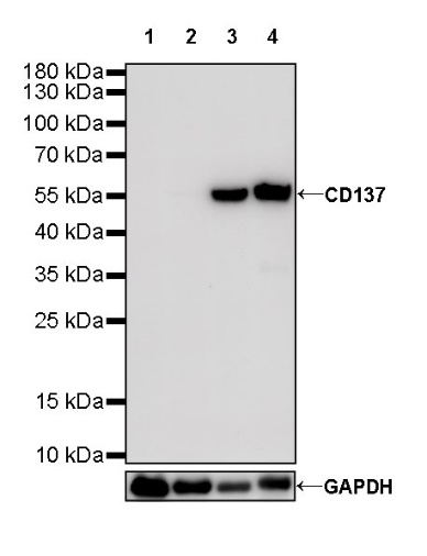
WB result of CD137 Rabbit mAb
Primary antibody: CD137 Rabbit mAb at 1/2000 dilution
Lane 1: mouse skeletal muscle lysate 20 µg
Lane 2: rat skeletal muscle lysate 20 µg
Lane 3: mouse thymus lysate 20 µg
Lane 4: rat thymus lysate 20 µg
Negative control: mouse skeletal muscle lysate; rat skeletal muscle lysate
Secondary antibody: Goat Anti-Rabbit IgG, (H+L), HRP conjugated at 1/10000 dilution
Predicted MW: 28 kDa
Observed MW: 55 kDa
Exposure time: 90 s
CD137 Rabbit mAb at 1/25 dilution (0.4 µg) immunoprecipitating CD137 in 0.4 mg mouse thymus lysate.
Western blot was performed on the immunoprecipitate using CD137 Rabbit mAb at 1/1000 dilution.
Secondary antibody (HRP) for IP was used at 1/1000 dilution.
Lane 1: mouse thymus lysate 5 µg (Input)
Lane 2: CD137 Rabbit mAb IP in mouse thymus lysate
Lane 3: Rabbit monoclonal IgG IP in mouse thymus lysate
Predicted MW: 28 kDa
Observed MW: 55 kDa
IHC shows positive staining in paraffin-embedded mouse thymus. Anti-CD137 antibody was used at 1/2000 dilution, followed by a HRP Polymer for Mouse & Rabbit IgG (ready to use). Counterstained with hematoxylin. Heat mediated antigen retrieval with Tris/EDTA buffer pH9.0 was performed before commencing with IHC staining protocol.
IHC shows positive staining in paraffin-embedded mouse liver. Anti-CD137 antibody was used at 1/2000 dilution, followed by a HRP Polymer for Mouse & Rabbit IgG (ready to use). Counterstained with hematoxylin. Heat mediated antigen retrieval with Tris/EDTA buffer pH9.0 was performed before commencing with IHC staining protocol.
IHC shows positive staining in paraffin-embedded rat thymus. Anti-CD137 antibody was used at 1/2000 dilution, followed by a HRP Polymer for Mouse & Rabbit IgG (ready to use). Counterstained with hematoxylin. Heat mediated antigen retrieval with Tris/EDTA buffer pH9.0 was performed before commencing with IHC staining protocol.
IHC shows positive staining in paraffin-embedded rat spleen. Anti-CD137 antibody was used at 1/2000 dilution, followed by a HRP Polymer for Mouse & Rabbit IgG (ready to use). Counterstained with hematoxylin. Heat mediated antigen retrieval with Tris/EDTA buffer pH9.0 was performed before commencing with IHC staining protocol.
 您现在的位置: