12 months from date of receipt / reconstitution, -20 °C as supplied
| 应用 | 稀释度 |
|---|---|
| WB | 1:1000 |
| FCM | 1:2000 |
| ICC | 1:500 |
Aminopeptidase N (also called CD13) is located in the small-intestinal and renal microvillar membrane, and also in other plasma membranes. In the small intestine aminopeptidase N plays a role in the final digestion of peptides generated from hydrolysis of proteins by gastric and pancreatic proteases. Its function in proximal tubular epithelial cells and other cell types is less clear. The large extracellular carboxyterminal domain contains a pentapeptide consensus sequence characteristic of members of the zinc-binding metalloproteinase superfamily. CD13 is also used by some viruses as a receptor to which these viruses bind to and then enter cells. It is a receptor for human coronavirus 229E, feline coronavirus serotype II (FCoV-II), TGEV, PEDV, canine coronavirus genotype II (CCoV-II) as well as several Deltacoronaviruses.
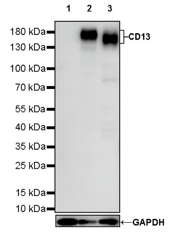
WB result of CD13 Mouse mAb
Primary antibody: CD13 Mouse mAb at 1/1000 dilution
Lane 1: HEK-293 whole cell lysate 20 µg
Lane 2: THP-1 whole cell lysate 20 µg
Lane 3: PANC-1 whole cell lysate 20 µg
Negative control: HEK-293 whole cell lysate
Secondary antibody: Goat Anti-Mouse IgG, (H+L), HRP conjugated at 1/10000 dilution
Predicted MW: 110 kDa
Observed MW: 150~160 kDa
Flow cytometric analysis of HEK293 (Human embryonic kidney epithelial cell, left) / THP-1 (Human monocytic leukemia monocyte, Right) cells labelling CD13 antibody at 1/2000 dilution (0.1 μg) / (Red) compared with a Mouse monoclonal IgG (Black) isotype control and an unlabelled control (cells without incubation with primary antibody and secondary antibody) (Blue). Goat Anti - Mouse IgG Alexa Fluor® 488 was used as the secondary antibody.
Negative control: HEK293
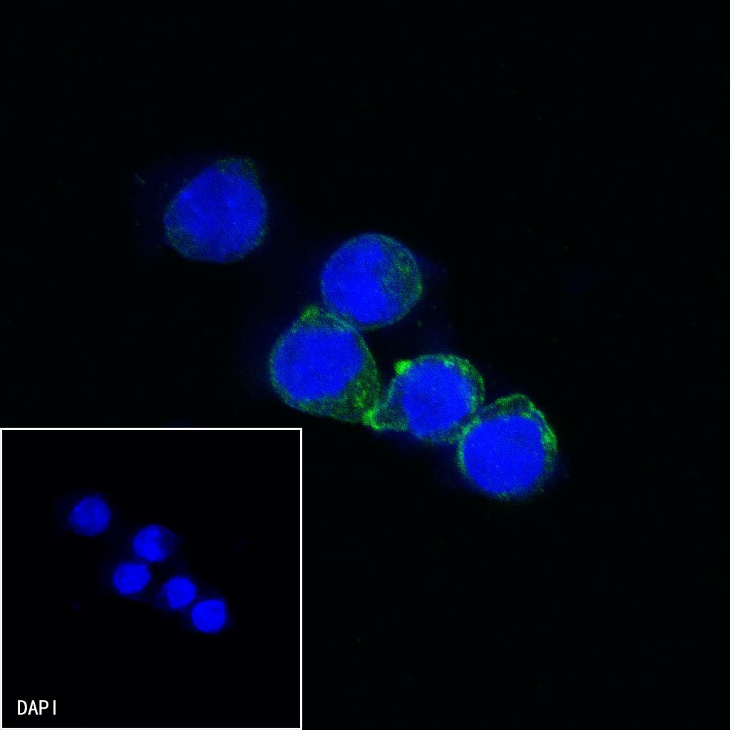
ICC shows positive staining in THP-1 cells. Anti-CD13 antibody was used at 1/500 dilution (Green) and incubated overnight at 4°C. Goat polyclonal Antibody to Mouse IgG - H&L (Alexa Fluor® 488) was used as secondary antibody at 1/1000 dilution. The cells were fixed with 100% ice-cold methanol and permeabilized with 0.1% PBS-Triton X-100. Nuclei were counterstained with DAPI (Blue).
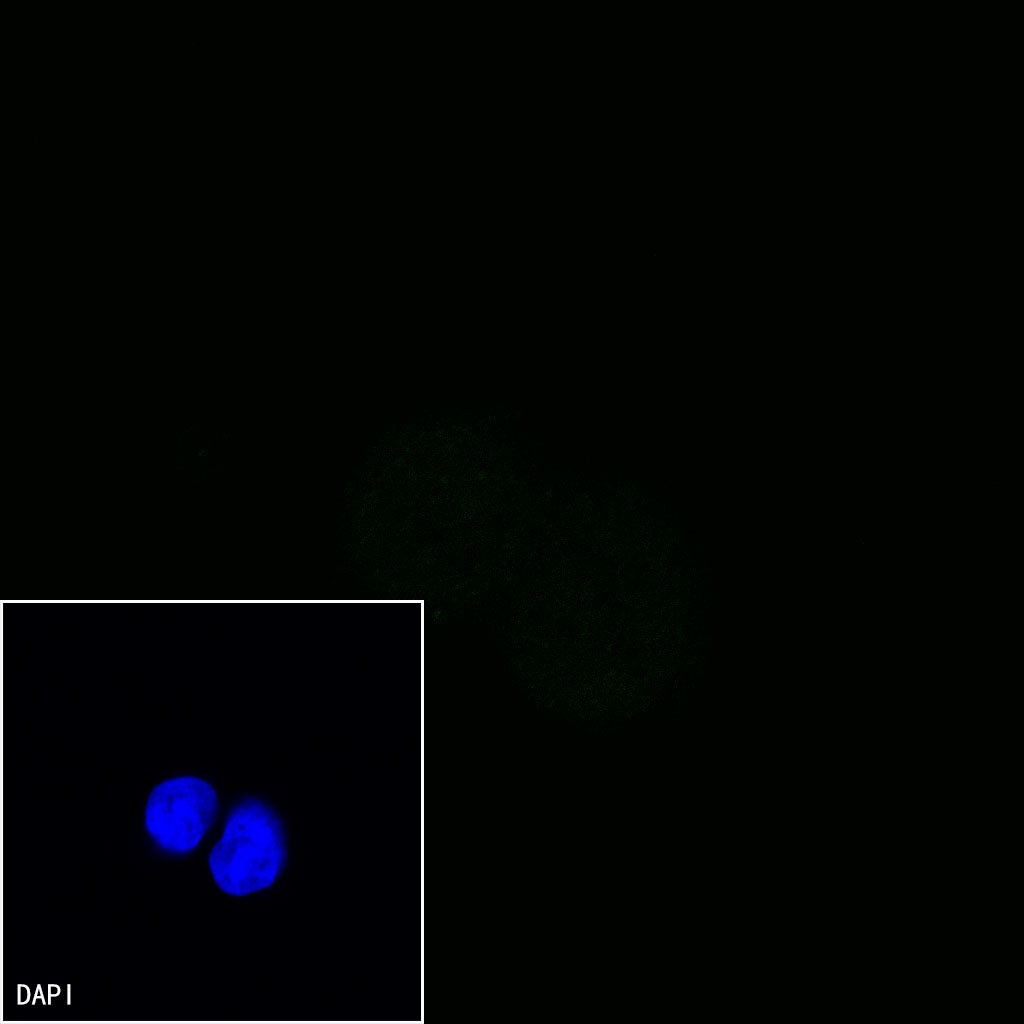
Negative control:ICC shows negative staining in HEK293 cells. Anti-CD13 antibody was used at 1/500 dilution and incubated overnight at 4°C. Goat polyclonal Antibody to Mouse IgG - H&L (Alexa Fluor® 488) was used as secondary antibody at 1/1000 dilution. The cells were fixed with 100% ice-cold methanol and permeabilized with 0.1% PBS-Triton X-100. Nuclei were counterstained with DAPI (Blue).
 您现在的位置: