
12 months from date of receipt / reconstitution, -20 °C as supplied
| 应用 | 稀释度 |
|---|---|
| WB | 1:1000 |
| ICC | 1:500 |
| ICFCM | 1:50 |
CCR7 is a transmembrane protein with 7 transmembrane domains, which is coupled with heterotrimeric G proteins, which transduce the signal downstream through various signaling cascades. The main function of the receptor is to guide immune cells to immune organs (lymph nodes, thymus, spleen) by detecting specific chemokines, which these tissues secrete. The receptor is expressed mostly on adaptive immune cell types, namely thymocytes, naive T and B cells, regulatory T cells, central memory lymphocytes, but also dendritic cells. CCR7 has been shown to stimulate dendritic cell maturation. CCR7 is also involved in homing of T cells to various secondary lymphoid organs such as lymph nodes and the spleen as well as trafficking of T cells within the spleen.
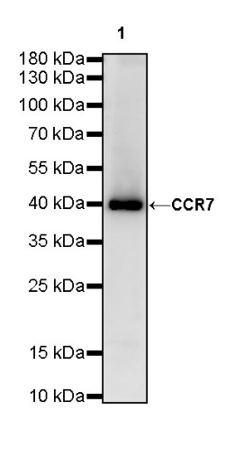
WB result of CCR7 Rabbit mAb
Primary antibody: CCR7 Rabbit mAb at 1/1000 dilution
Lane 1: K562 whole cell lysate 20 µg
Secondary antibody: Goat Anti-Rabbit IgG, (H+L), HRP conjugated at 1/10000 dilution
Predicted MW: 43 kDa
Observed MW: 39 kDa
(This blot was developed with high sensitivity substrate)
WB result of CCR7 Rabbit mAb
Primary antibody: CCR7 Rabbit mAb at 1/1000 dilution
Lane 1: mouse spleen lysate 20 µg
Lane 2: mouse lung lysate 20 µg
Secondary antibody: Goat Anti-Rabbit IgG, (H+L), HRP conjugated at 1/10000 dilution
Predicted MW: 43 kDa
Observed MW: 39 kDa
(This blot was developed with high sensitivity substrate)
WB result of CCR7 Rabbit mAb
Primary antibody: CCR7 Rabbit mAb at 1/1000 dilution
Lane 1: rat spleen lysate 20 µg
Lane 2: rat lung lysate 20 µg
Secondary antibody: Goat Anti-Rabbit IgG, (H+L), HRP conjugated at 1/10000 dilution
Predicted MW: 43 kDa
Observed MW: 39 kDa
(This blot was developed with high sensitivity substrate)
Flow cytometric analysis of 4% PFA fixed 90% methanol permeabilized Jurkat (Human T cell leukemia T lymphocyte) labelling CCR7 antibody at 1/50 dilution (1 μg) / (Red) compared with a Rabbit monoclonal IgG (Black) isotype control and an unlabelled control (cells without incubation with primary antibody and secondary antibody) (Blue). Goat Anti - Rabbit IgG Alexa Fluor® 488 was used as the secondary antibody.
ICC shows positive staining in Jurkat cells. Anti-CCR7 antibody was used at 1/500 dilution (Green) and incubated overnight at 4°C. Goat polyclonal Antibody to Rabbit IgG - H&L (Alexa Fluor® 488) was used as secondary antibody at 1/1000 dilution. The cells were fixed with 100% ice-cold methanol and permeabilized with 0.1% PBS-Triton X-100. Nuclei were counterstained with DAPI (Blue). Counterstain with tubulin (Red).
 您现在的位置: