
12 months from date of receipt / reconstitution, -20 °C as supplied
| 应用 | 稀释度 |
|---|---|
| WB | 1:1000 |
| IHC-P | 1:2000 |
| IP | 1:50 |
Paxillin is a protein that in humans is encoded by the PXN gene. Paxillin is expressed at focal adhesions of non-striated cells and at costameres of striated muscle cells, and it functions to adhere cells to the extracellular matrix. Paxillin has been shown to have a clinically-significant role in patients with several cancer types. Enhanced expression of paxillin has been detected in premalignant areas of hyperplasia, squamous metaplasia and goblet cell metaplasia, as well as dysplastic lesions and carcinoma in high-risk patients with lung adenocarcinoma. Mutations in PXN have been associated with enhanced tumor growth, cell proliferation, and invasion in lung cancer tissues.
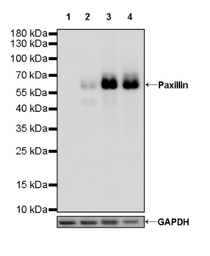
WB result of Paxillin Rabbit mAb
Primary antibody: Paxillin Rabbit mAb at 1/1000 dilution
Lane 1: HL-60 whole cell lysate 5 µg
Lane 2: HeLa whole cell lysate 5 µg
Lane 3: U-87 MG whole cell lysate 5 µg
Lane 4: PC-3 whole cell lysate 5 µg
Negative control: HL-60 whole cell lysate
Secondary antibody: Goat Anti-Rabbit IgG, (H+L), HRP conjugated at 1/10000 dilution
Predicted MW: 65 kDa
Observed MW: 65 kDa
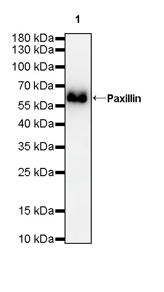
WB result of Paxillin Rabbit mAb
Primary antibody: Paxillin Rabbit mAb at 1/1000 dilution
Lane 1: C6 whole cell lysate 5 µg
Secondary antibody: Goat Anti-Rabbit IgG, (H+L), HRP conjugated at 1/10000 dilution
Predicted MW: 65 kDa
Observed MW: 65 kDa
WB result of Paxillin Rabbit mAb
Primary antibody: Paxillin Rabbit mAb at 1/1000 dilution
Lane 1: RAW 264.7 whole cell lysate 5 µg
Secondary antibody: Goat Anti-Rabbit IgG, (H+L), HRP conjugated at 1/10000 dilution
Predicted MW: 65 kDa
Observed MW: 65 kDa
(This blot was developed with high sensitivity substrate)
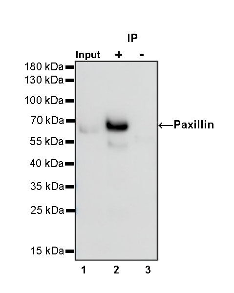
Paxillin Rabbit mAb at 1/50 dilution (1 µg) immunoprecipitating Paxillin in 0.4 mg U-87 MG whole cell lysate.
Western blot was performed on the immunoprecipitate using Paxillin Rabbit mAb at 1/1000 dilution.
Secondary antibody (HRP) for IP was used at 1/400 dilution.
Lane 1: U-87 MG whole cell lysate 5 µg (Input)
Lane 2: Paxillin Rabbit mAb IP in U-87 MG whole cell lysate
Lane 3: Rabbit monoclonal IgG IP in U-87 MG whole cell lysate
Predicted MW: 65 kDa
Observed MW: 65 kDa
IHC shows positive staining in paraffin-embedded human placenta. Anti-Paxillin antibody was used at 1/2000 dilution, followed by a HRP Polymer for Mouse & Rabbit IgG (ready to use). Counterstained with hematoxylin. Heat mediated antigen retrieval with Tris/EDTA buffer pH9.0 was performed before commencing with IHC staining protocol.
IHC shows positive staining in paraffin-embedded human tonsil. Anti-Paxillin antibody was used at 1/2000 dilution, followed by a HRP Polymer for Mouse & Rabbit IgG (ready to use). Counterstained with hematoxylin. Heat mediated antigen retrieval with Tris/EDTA buffer pH9.0 was performed before commencing with IHC staining protocol.
IHC shows positive staining in paraffin-embedded human prostate. Anti-Paxillin antibody was used at 1/2000 dilution, followed by a HRP Polymer for Mouse & Rabbit IgG (ready to use). Counterstained with hematoxylin. Heat mediated antigen retrieval with Tris/EDTA buffer pH9.0 was performed before commencing with IHC staining protocol.
IHC shows positive staining in paraffin-embedded human skeletal muscle. Anti-Paxillin antibody was used at 1/2000 dilution, followed by a HRP Polymer for Mouse & Rabbit IgG (ready to use). Counterstained with hematoxylin. Heat mediated antigen retrieval with Tris/EDTA buffer pH9.0 was performed before commencing with IHC staining protocol.
IHC shows positive staining in paraffin-embedded human breast cancer. Anti-Paxillin antibody was used at 1/2000 dilution, followed by a HRP Polymer for Mouse & Rabbit IgG (ready to use). Counterstained with hematoxylin. Heat mediated antigen retrieval with Tris/EDTA buffer pH9.0 was performed before commencing with IHC staining protocol.
IHC shows positive staining in paraffin-embedded human prostatic cancer. Anti-Paxillin antibody was used at 1/2000 dilution, followed by a HRP Polymer for Mouse & Rabbit IgG (ready to use). Counterstained with hematoxylin. Heat mediated antigen retrieval with Tris/EDTA buffer pH9.0 was performed before commencing with IHC staining protocol.
IHC shows positive staining in paraffin-embedded mouse liver. Anti-Paxillin antibody was used at 1/2000 dilution, followed by a HRP Polymer for Mouse & Rabbit IgG (ready to use). Counterstained with hematoxylin. Heat mediated antigen retrieval with Tris/EDTA buffer pH9.0 was performed before commencing with IHC staining protocol.
IHC shows positive staining in paraffin-embedded rat cardiac muscle. Anti-Paxillin antibody was used at 1/2000 dilution, followed by a HRP Polymer for Mouse & Rabbit IgG (ready to use). Counterstained with hematoxylin. Heat mediated antigen retrieval with Tris/EDTA buffer pH9.0 was performed before commencing with IHC staining protocol.
 您现在的位置: