
12 months from date of receipt / reconstitution, -20 °C as supplied
| 应用 | 稀释度 |
|---|---|
| WB | 1:1000 |
| IP | 1:50 |
| IHC-P | 1:500 |
P27 is a protein which belongs to the Cip/Kip family of cyclin dependent kinase (Cdk) inhibitor proteins. It binds to and prevents the activation of cyclin E-CDK2 or cyclin D-CDK4 complexes, and thus controls the cell cycle progression at G1. It is often referred to as a cell cycle inhibitor protein because its major function is to stop or slow down the cell division cycle. p27 is considered a tumor suppressor because of its function as a regulator of the cell cycle. In cancers it is often inactivated via impaired synthesis, accelerated degradation, or mislocalization. p27 can also be mislocalized to the cytoplasm in order to facilitate metastasis.
WB result of p27 Rabbit mAb
Primary antibody: p27 Rabbit mAb at 1/1000 dilution
Lane 1: NIH/3T3 whole cell lysate 20 µg
Lane 2: Raw 264.7 whole cell lysate 20 µg
Lane 3: mouse thymus lysate 20 µg
Secondary antibody: Goat Anti-Rabbit IgG, (H+L), HRP conjugated at 1/10000 dilution
Predicted MW: 22 kDa
Observed MW: 24 kDa
WB result of p27 Rabbit mAb
Primary antibody: p27 Rabbit mAb at 1/1000 dilution
Lane 1: C6 whole cell lysate 20 µg
Lane 2: rat thymus lysate 20 µg
Secondary antibody: Goat Anti-Rabbit IgG, (H+L), HRP conjugated at 1/10000 dilution
Predicted MW: 22 kDa
Observed MW: 25 kDa
p27 Rabbit mAb at 1/50 dilution (1 µg) immunoprecipitating p27 in 0.4 mg NIH/3T3 whole cell lysate.
Western blot was performed on the immunoprecipitate using p27 Rabbit mAb at 1/1000 dilution.
Secondary antibody (HRP) for IP was used at 1/400 dilution.
Lane 1: NIH/3T3 whole cell lysate 20 µg (Input)
Lane 2: p27 Rabbit mAb IP in NIH/3T3 whole cell lysate
Lane 3: Rabbit monoclonal IgG IP in NIH/3T3 whole cell lysate
Predicted MW: 22 kDa
Observed MW: 25 kDa
IHC shows positive staining in paraffin-embedded rat testis. Anti-p27 antibody was used at 1/500 dilution, followed by a HRP Polymer for Mouse & Rabbit IgG (ready to use). Counterstained with hematoxylin. Heat mediated antigen retrieval with Tris/EDTA buffer pH9.0 was performed before commencing with IHC staining protocol.
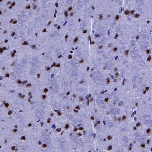
IHC shows positive staining in paraffin-embedded mouse cerebral cortex. Anti-p27 antibody was used at 1/500 dilution, followed by a HRP Polymer for Mouse & Rabbit IgG (ready to use). Counterstained with hematoxylin. Heat mediated antigen retrieval with Tris/EDTA buffer pH9.0 was performed before commencing with IHC staining protocol.
IHC shows positive staining in paraffin-embedded mouse thymus. Anti-p27 antibody was used at 1/500 dilution, followed by a HRP Polymer for Mouse & Rabbit IgG (ready to use). Counterstained with hematoxylin. Heat mediated antigen retrieval with Tris/EDTA buffer pH9.0 was performed before commencing with IHC staining protocol.
IHC shows positive staining in paraffin-embedded mouse lung. Anti-p27 antibody was used at 1/500 dilution, followed by a HRP Polymer for Mouse & Rabbit IgG (ready to use). Counterstained with hematoxylin. Heat mediated antigen retrieval with Tris/EDTA buffer pH9.0 was performed before commencing with IHC staining protocol.
IHC shows positive staining in paraffin-embedded mouse testis. Anti-p27 antibody was used at 1/500 dilution, followed by a HRP Polymer for Mouse & Rabbit IgG (ready to use). Counterstained with hematoxylin. Heat mediated antigen retrieval with Tris/EDTA buffer pH9.0 was performed before commencing with IHC staining protocol.
IHC shows positive staining in paraffin-embedded rat kidney. Anti-p27 antibody was used at 1/500 dilution, followed by a HRP Polymer for Mouse & Rabbit IgG (ready to use). Counterstained with hematoxylin. Heat mediated antigen retrieval with Tris/EDTA buffer pH9.0 was performed before commencing with IHC staining protocol.
 您现在的位置: