
12 months from date of receipt / reconstitution, -20 °C as supplied
| 应用 | 稀释度 |
|---|---|
| IHC-P | 1:2000 |
The LAG3 protein, which belongs to immunoglobulin (Ig) superfamily, comprises a 503-amino acid type I transmembrane protein with four extracellular Ig-like domains, designated D1 to D4. LAG-3 is expressed on activated T cells, natural killer cells, B cells and plasmacytoid dendritic cells. LAG3's main ligand is MHC class II, to which it binds with higher affinity than CD4. The protein negatively regulates cellular proliferation, activation, and homeostasis of T cells, in a similar fashion to CTLA-4 and PD-1 and has been reported to play a role in Treg suppressive function. Fibrinogen-like protein1 FGL1, a liver-secreted protein, is another (major) LAG3 functional ligand independent of MHC-II. LAG3 also helps maintain CD8+ T cells in a tolerogenic state and, working with PD-1, helps maintain CD8 exhaustion during chronic viral infection. LAG3 is known to be involved in the maturation and activation of dendritic cells.
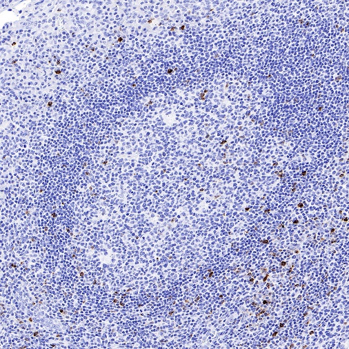
IHC shows positive staining in paraffin-embedded human tonsil. Anti-LAG3 antibody was used at 1/2000 dilution, followed by a HRP Polymer for Mouse & Rabbit IgG (ready to use). Counterstained with hematoxylin. Heat mediated antigen retrieval with Tris/EDTA buffer pH9.0 was performed before commencing with IHC staining protocol.
IHC shows positive staining in paraffin-embedded human breast cancer. Anti-LAG3 antibody was used at 1/2000 dilution, followed by a HRP Polymer for Mouse & Rabbit IgG (ready to use). Counterstained with hematoxylin. Heat mediated antigen retrieval with Tris/EDTA buffer pH9.0 was performed before commencing with IHC staining protocol.
IHC shows positive staining in paraffin-embedded human colon cancer. Anti-LAG3 antibody was used at 1/2000 dilution, followed by a HRP Polymer for Mouse & Rabbit IgG (ready to use). Counterstained with hematoxylin. Heat mediated antigen retrieval with Tris/EDTA buffer pH9.0 was performed before commencing with IHC staining protocol.
IHC shows positive staining in paraffin-embedded human Hodgkin’s lymphoma. Anti-LAG3 antibody was used at 1/2000 dilution, followed by a HRP Polymer for Mouse & Rabbit IgG (ready to use). Counterstained with hematoxylin. Heat mediated antigen retrieval with Tris/EDTA buffer pH9.0 was performed before commencing with IHC staining protocol.
Negative control: IHC shows negative staining in paraffin-embedded human mantle lymphoma. Anti-LAG3 antibody was used at 1/2000 dilution, followed by a HRP Polymer for Mouse & Rabbit IgG (ready to use). Counterstained with hematoxylin. Heat mediated antigen retrieval with Tris/EDTA buffer pH9.0 was performed before commencing with IHC staining protocol.
Negative control: IHC shows negative staining in paraffin-embedded human diffuse large B-cell lymphoma. Anti-LAG3 antibody was used at 1/2000 dilution, followed by a HRP Polymer for Mouse & Rabbit IgG (ready to use). Counterstained with hematoxylin. Heat mediated antigen retrieval with Tris/EDTA buffer pH9.0 was performed before commencing with IHC staining protocol.
 您现在的位置: