
12 months from date of receipt / reconstitution, -20 °C as supplied
| 应用 | 稀释度 |
|---|---|
| WB | 1:1000 |
| IP | 1:50 |
| IHC-P | 1:2000 |
SOX1 is a gene that encodes a transcription factor with a HMG-box (high mobility group) DNA-binding domain and functions primarily in neurogenesis. SOX1 exerts its importance in its role in development of the central nervous system (neurogenesis) and in particular the development of the eye, where it is functionally redundant with SOX3 and to a lesser degree SOX2, and maintenance of neural progenitor cell identity. SOX1 expression is restricted to the neuroectoderm by proliferating progenitor cells in the tetrapod embryo.
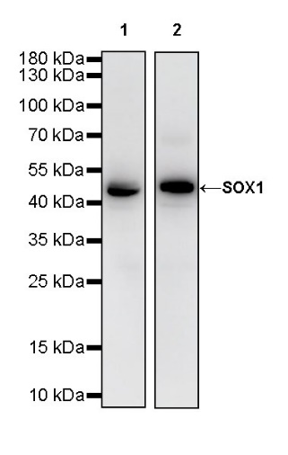
WB result of SOX1 Rabbit mAb
Primary antibody: SOX1 Rabbit mAb at 1/1000 dilution
Lane 1: Neuro-2a lysate 20 µg
Lane 2: mouse brain lysate 20 µg
Secondary antibody: Goat Anti-Rabbit IgG, (H+L), HRP conjugated at 1/10000 dilution
Predicted MW: 39 kDa
Observed MW: 43kDa
(This blot was developed with high sensitivity substrate)
WB result of SOX1 Rabbit mAb
Primary antibody: SOX1 Rabbit mAb at 1/1000 dilution
Lane 1: rat brain lysate 20 µg
Secondary antibody: Goat Anti-Rabbit IgG, (H+L), HRP conjugated at 1/10000 dilution
Predicted MW: 39 kDa
Observed MW: 43 kDa
(This blot was developed with high sensitivity substrate)
SOX1 Rabbit mAb at 1/50 dilution (1 µg) immunoprecipitating SOX1 in 0.4 mg mouse brain lysate.
Western blot was performed on the immunoprecipitate using SOX1 Rabbit mAb at 1/1000 dilution.
Secondary antibody (HRP) for IP was used at 1/400 dilution.
Lane 1: mouse brain lysate 20 µg (Input)
Lane 2: SOX1 Rabbit mAb IP in mouse brain lysate
Lane 3: Rabbit monoclonal IgG IP in mouse brain lysate
Predicted MW: 39 kDa
Observed MW: 43 kDa
(This blot was developed with high sensitivity substrate)
IHC shows positive staining in paraffin-embedded mouse cerebral cortex. Anti-SOX1 antibody was used at 1/2000 dilution, followed by a HRP Polymer for Mouse & Rabbit IgG (ready to use). Counterstained with hematoxylin. Heat mediated antigen retrieval with Tris/EDTA buffer pH9.0 was performed before commencing with IHC staining protocol.
IHC shows positive staining in paraffin-embedded mouse cerebellum. Anti-SOX1 antibody was used at 1/2000 dilution, followed by a HRP Polymer for Mouse & Rabbit IgG (ready to use). Counterstained with hematoxylin. Heat mediated antigen retrieval with Tris/EDTA buffer pH9.0 was performed before commencing with IHC staining protocol.
IHC shows positive staining in paraffin-embedded rat cerebral cortex. Anti-SOX1 antibody was used at 1/2000 dilution, followed by a HRP Polymer for Mouse & Rabbit IgG (ready to use). Counterstained with hematoxylin. Heat mediated antigen retrieval with Tris/EDTA buffer pH9.0 was performed before commencing with IHC staining protocol.
IHC shows positive staining in paraffin-embedded rat cerebellum. Anti-SOX1 antibody was used at 1/2000 dilution, followed by a HRP Polymer for Mouse & Rabbit IgG (ready to use). Counterstained with hematoxylin. Heat mediated antigen retrieval with Tris/EDTA buffer pH9.0 was performed before commencing with IHC staining protocol.
 您现在的位置: