
12 months from date of receipt / reconstitution, -20 °C as supplied
| 应用 | 稀释度 |
|---|---|
| ICC | 1:500 |
| FCM | 1:10000 |
CD45 is a member of the protein tyrosine phosphatase (PTP) family. CD45 is a type I transmembrane protein that is present in various isoforms on all differentiated hematopoietic cells (except erythrocytes and plasma cells). CD45 has been shown to be an essential regulator of T- and B-cell antigen receptor signaling. It functions through either direct interaction with components of the antigen receptor complexes via its extracellular domain (a form of co-stimulation), or by activating various Src family kinases required for the antigen receptor signaling via its cytoplasmic domain. CD45 also suppresses JAK kinases, and so functions as a negative regulator of cytokine receptor signaling.
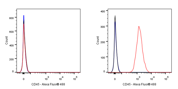
Flow cytometric analysis of HeLa (Human cervix adenocarcinoma epithelial cell, left) / Jurkat (Human T cell leukemia T lymphocyte, Right) cells labelling CD45 antibody at 1/10000 dilution (0.01 μg) / (Red) compared with a Mouse monoclonal IgG (Black) isotype control and an unlabelled control (cells without incubation with primary antibody and secondary antibody) (Blue). Goat Anti - Mouse IgG Alexa Fluor® 488 was used as the secondary antibody.
Negative control: HeLa
Flow cytometric analysis of human PBMC (human peripheral blood mononuclear cell) labelling CD45 antibody at 1/1000 dilution (0.1 μg) / (Red) compared with a Mouse monoclonal IgG (Black) isotype control and an unlabelled control (cells without incubation with primary antibody and secondary antibody) (Blue). Goat Anti - Mouse IgG Alexa Fluor® 488 was used as the secondary antibody.
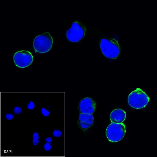
ICC shows positive staining in Jurkat cells. Anti-CD45 antibody was used at 1/500 dilution (Green) and incubated overnight at 4°C. Goat polyclonal Antibody to Mouse IgG - H&L (Alexa Fluor® 488) was used as secondary antibody at 1/1000 dilution. The cells were fixed with 100% ice-cold methanol and permeabilized with 0.1% PBS-Triton X-100. Nuclei were counterstained with DAPI (Blue).
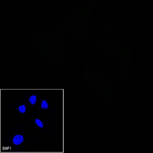
Negative control: ICC shows negative staining in HeLa cells. Anti-CD45 antibody was used at 1/500 dilution and incubated overnight at 4°C. Goat polyclonal Antibody to Mouse IgG - H&L (Alexa Fluor® 488) was used as secondary antibody at 1/1000 dilution. The cells were fixed with 100% ice-cold methanol and permeabilized with 0.1% PBS-Triton X-100. Nuclei were counterstained with DAPI (Blue).
 您现在的位置:
