
12 months from date of receipt / reconstitution, -20 °C as supplied
| 应用 | 稀释度 |
|---|---|
| WB | 1:1000 |
| IP | 1:50 |
| IHC-P | 1:100 |
WW domain-containing proteins are found in all eukaryotes and play an important role in the regulation of a wide variety of cellular functions such as protein degradation, transcription, and RNA splicing. The highest normal expression of WWOX is detected in hormonally regulated tissues such as testis, ovary, and prostate. This expression pattern and the presence of an SRD domain suggest a role for this protein in steroid metabolism. WWOX is more than 90% identical to the mouse protein, which is an essential mediator of tumor necrosis factor-alpha-induced apoptosis, suggesting a similar, important role in apoptosis for the human protein. In addition, there is evidence that this protein behaves as a suppressor of tumor growth.
WB result of WWOX Rabbit mAb
Primary antibody: WWOX Rabbit mAb at 1/1000 dilution
Lane 1: mouse testis lysate 20 µg
Secondary antibody: Goat Anti-Rabbit IgG, (H+L), HRP conjugated at 1/10000 dilution
Predicted MW: 47 kDa
Observed MW: 46 kDa
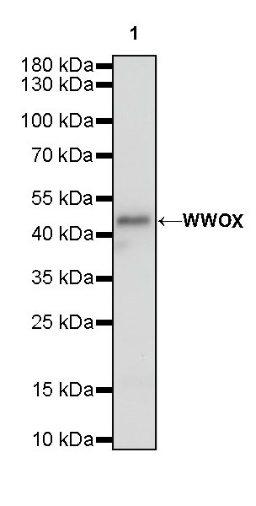
WB result of WWOX Rabbit mAb
Primary antibody: WWOX Rabbit mAb at 1/1000 dilution
Lane 1: rat brain lysate 20 µg
Secondary antibody: Goat Anti-Rabbit IgG, (H+L), HRP conjugated at 1/10000 dilution
Predicted MW: 47 kDa
Observed MW: 46 kDa
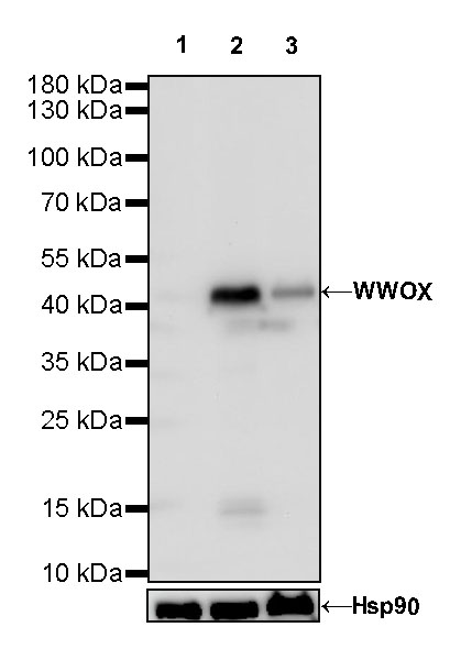
WB result of WWOX Rabbit mAb
Primary antibody: WWOX Rabbit mAb at 1/1000 dilution
Lane 1: MDA-MB-231 whole cell lysate 20 µg
Lane 2: MCF7 whole cell lysate 20 µg
Lane 3: HepG2 whole cell lysate 20 µg
Negative control: MDA-MB-231 whole cell lysate
Secondary antibody: Goat Anti-Rabbit IgG, (H+L), HRP conjugated at 1/10000 dilution
Predicted MW: 47 kDa
Observed MW: 46 kDa
WWOX Rabbit mAb at 1/50 dilution (1 µg) immunoprecipitating WWOX in 0.4 mg MCF7 whole cell lysate.
Western blot was performed on the immunoprecipitate using WWOX Rabbit mAb at 1/1000 dilution.
Secondary antibody (HRP) for IP was used at 1/400 dilution.
Lane 1: MCF7 whole cell lysate 20 µg (Input)
Lane 2: WWOX Rabbit mAb IP in MCF7 whole cell lysate
Lane 3: Rabbit monoclonal IgG IP in MCF7 whole cell lysate
Predicted MW: 47 kDa
Observed MW: 44 kDa
IHC shows positive staining in paraffin-embedded human kidney. Anti-WWOX antibody was used at 1/100 dilution, followed by a HRP Polymer for Mouse & Rabbit IgG (ready to use). Counterstained with hematoxylin. Heat mediated antigen retrieval with Tris/EDTA buffer pH9.0 was performed before commencing with IHC staining protocol.
IHC shows positive staining in paraffin-embedded human breast cancer. Anti-WWOX antibody was used at 1/100 dilution, followed by a HRP Polymer for Mouse & Rabbit IgG (ready to use). Counterstained with hematoxylin. Heat mediated antigen retrieval with Tris/EDTA buffer pH9.0 was performed before commencing with IHC staining protocol.
IHC shows positive staining in paraffin-embedded human hepatocellular carcinoma. Anti-WWOX antibody was used at 1/100 dilution, followed by a HRP Polymer for Mouse & Rabbit IgG (ready to use). Counterstained with hematoxylin. Heat mediated antigen retrieval with Tris/EDTA buffer pH9.0 was performed before commencing with IHC staining protocol.
IHC shows positive staining in paraffin-embedded rat liver. Anti-WWOX antibody was used at 1/100 dilution, followed by a HRP Polymer for Mouse & Rabbit IgG (ready to use). Counterstained with hematoxylin. Heat mediated antigen retrieval with Tris/EDTA buffer pH9.0 was performed before commencing with IHC staining protocol.
 您现在的位置: