
12 months from date of receipt / reconstitution, -20 °C as supplied
| 应用 | 稀释度 |
|---|---|
| WB | 1:1000 |
| ICC | 1:500 |
| IP | 1:50 |
GSPT1 is a protein involved in the termination of translation, a process in which ribosomes synthesize proteins after the transcription of DNA to RNA. There is a demonstrated link between GSPT1 degradation and antitumor activity.
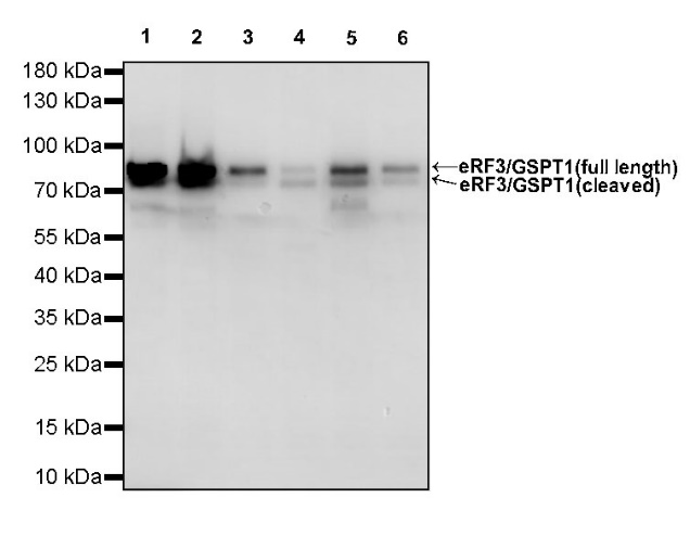
WB result of eRF3/GSPT1 Rabbit mAb
Primary antibody: eRF3/GSPT1 Rabbit mAb at 1/1000 dilution
Lane 1: HeLa whole cell lysate 20 µg
Lane 2: 293T whole cell lysate 20 µg
Lane 3: PC-3 whole cell lysate 20 µg
Lane 4: A431 whole cell lysate 20 µg
Lane 5: HepG2 whole cell lysate 20 µg
Lane 6: SK-OV-3 whole cell lysate 20 µg
Secondary antibody: Goat Anti-Rabbit IgG, (H+L), HRP conjugated at 1/10000 dilution
Predicted MW: 55 kDa
Observed MW: 80 kDa
WB result of eRF3/GSPT1 Rabbit mAb
Primary antibody: eRF3/GSPT1 Rabbit mAb at 1/1000 dilution
Lane 1: NIH/3T3 whole cell lysate 20 µg
Lane 2: Raw 264.7 whole cell lysate 20 µg
Lane 3: mouse brain lysate 20 µg
Lane 4: mouse testis lysate 20 µg
Lane 5: mouse spleen lysate 20 µg
Secondary antibody: Goat Anti-Rabbit IgG, (H+L), HRP conjugated at 1/10000 dilution
Predicted MW: 55 kDa
Observed MW: 80 kDa
WB result of eRF3/GSPT1 Rabbit mAb
Primary antibody: eRF3/GSPT1 Rabbit mAb at 1/1000 dilution
Lane 1: rat brain lysate 20 µg
Lane 2: rat testis lysate 20 µg
Lane 3: rat spleen lysate 20 µg
Secondary antibody: Goat Anti-Rabbit IgG, (H+L), HRP conjugated at 1/10000 dilution
Predicted MW: 55 kDa
Observed MW: 80 kDa
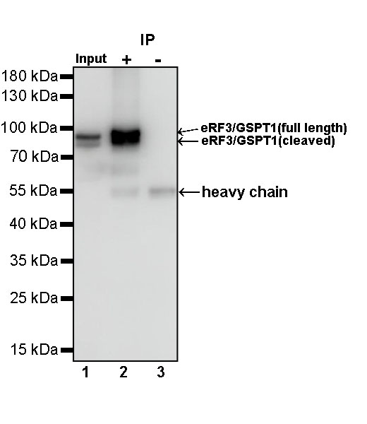
eRF3/GSPT1 Rabbit mAb at 1/50 dilution (1 µg) immunoprecipitating eRF3/GSPT1 in 0.4 mg PC-12 whole cell lysate.
Western blot was performed on the immunoprecipitate using eRF3/GSPT1 Rabbit mAb at 1/1000 dilution.
Secondary antibody (HRP) for IP was used at 1/400 dilution.
Lane 1: PC-12 whole cell lysate 20 µg (Input)
Lane 2: eRF3/GSPT1 Rabbit mAb IP in PC-12 whole cell lysate
Lane 3: Rabbit monoclonal IgG IP in PC-12 whole cell lysate
Predicted MW: 55 kDa
Observed MW: 80 kDa
ICC shows positive staining in PC-3 cells. Anti-eRF3 GSPT1 antibody was used at 1/500 dilution (Green) and incubated overnight at 4°C. Goat polyclonal Antibody to Rabbit IgG - H&L (Alexa Fluor® 488) was used as secondary antibody at 1/1000 dilution. The cells were fixed with 100% ice-cold methanol and permeabilized with 0.1% PBS-Triton X-100. Nuclei were counterstained with DAPI (Blue). Counterstain with tubulin (Red).
 您现在的位置: