
12 months from date of receipt / reconstitution, -20 °C as supplied
| 应用 | 稀释度 |
|---|---|
| WB | 1:1000 |
| IHC | 1:100-1:200 |
| ICC | 1:50 |
VCAM-1 is a member of the immunoglobulin superfamily, the superfamily of proteins including antibodies and T-cell receptors. It mediates the adhesion of lymphocytes, monocytes, eosinophils, and basophils to vascular endothelium. It also functions in leukocyte-endothelial cell signal transduction, and it may play a role in the development of atherosclerosis and rheumatoid arthritis. Certain melanoma cells can use VCAM-1 to adhere to the endothelium, VCAM-1 may participate in monocyte recruitment to atherosclerotic sites, and it is highly overexpressed in the inflamed brain. As a result, VCAM-1 is a potential drug target.
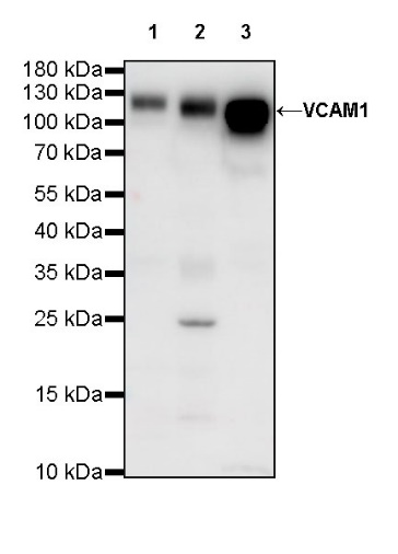
WB result of VCAM1 Rabbit mAb
Primary antibody: VCAM1 Rabbit mAb at 1/1000 dilution
Lane 1: HUVEC whole cell lysate 20 µg
Lane 2: HuT 78 whole cell lysate 20 µg
Lane 3: C2C12 whole cell lysate 20 µg
Secondary antibody: Goat Anti-Rabbit IgG, (H+L), HRP conjugated at 1/10000 dilution
Predicted MW: 81 kDa
Observed MW: 110~120 kDa
(This blot was developed with high sensitivity substrate)
WB result of VCAM1 Rabbit mAb
Primary antibody: VCAM1 Rabbit mAb at 1/1000 dilution
Lane 1: mouse spleen lysate 20 µg
Secondary antibody: Goat Anti-Rabbit IgG, (H+L), HRP conjugated at 1/10000 dilution
Predicted MW: 81 kDa
Observed MW: 110 kDa
(This blot was developed with high sensitivity substrate)
WB result of VCAM1 Rabbit mAb
Primary antibody: VCAM1 Rabbit mAb at 1/1000 dilution
Lane 1: rat spleen lysate 20 µg
Secondary antibody: Goat Anti-Rabbit IgG, (H+L), HRP conjugated at 1/10000 dilution
Predicted MW: 81 kDa
Observed MW: 110 kDa
(This blot was developed with high sensitivity substrate)
IHC shows positive staining in paraffin-embedded human tonsil. Anti-VCAM1 antibody was used at 1/100 dilution, followed by a HRP Polymer for Mouse & Rabbit IgG (ready to use). Counterstained with hematoxylin. Heat mediated antigen retrieval with Tris/EDTA buffer pH9.0 was performed before commencing with IHC staining protocol.
IHC shows positive staining in paraffin-embedded human colon. Anti-VCAM1 antibody was used at 1/200 dilution, followed by a HRP Polymer for Mouse & Rabbit IgG (ready to use). Counterstained with hematoxylin. Heat mediated antigen retrieval with Tris/EDTA buffer pH9.0 was performed before commencing with IHC staining protocol.
IHC shows positive staining in paraffin-embedded mouse spleen. Anti-VCAM1 antibody was used at 1/200 dilution, followed by a HRP Polymer for Mouse & Rabbit IgG (ready to use). Counterstained with hematoxylin. Heat mediated antigen retrieval with Tris/EDTA buffer pH9.0 was performed before commencing with IHC staining protocol.
ICC shows positive staining in C2C12 cells. Anti-VCAM1 antibody was used at 1/50 dilution (Green) and incubated overnight at 4°C. Goat polyclonal Antibody to Rabbit IgG - H&L (Alexa Fluor® 488) was used as secondary antibody at 1/1000 dilution. The cells were fixed with 100% ice-cold methanol and permeabilized with 0.1% PBS-Triton X-100. Nuclei were counterstained with DAPI (Blue). Counterstain with tubulin (Red).
 您现在的位置: