
12 months from date of receipt / reconstitution, -20 °C as supplied
| 应用 | 稀释度 |
|---|---|
| WB | 1:1000 |
| IHC | 1:500-1:2000 |
| ICC | 1:500 |
Cyclic GMP-AMP synthase (cGAS, cGAMP synthase), belonging to the nucleotidyltransferase family, is a cytosolic DNA sensor that activates a type-I interferon response. It is part of the cGAS-STING DNA sensing pathway. It binds to microbial DNA as well as self DNA that invades the cytoplasm, and catalyzes cGAMP synthesis. cGAMP then functions as a second messenger that binds to and activates the endoplasmic reticulum protein STING to trigger type-I IFNs production. Mice lacking cGAS are more vulnerable to lethal infection by DNA viruses and RNA viruses.[5][6] In addition, cGAS has been shown to be an innate immune sensor of retroviruses including HIV.
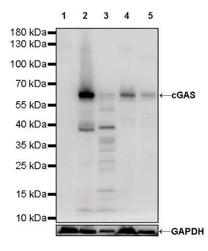
WB result of cGAS Rabbit mAb
Primary antibody: cGAS Rabbit mAb at 1/1000 dilution
Lane 1: 293T whole cell lysate 30 µg
Lane 2: MCF7 whole cell lysate 30 µg
Lane 3: THP-1 whole cell lysate 30 µg
Lane 4: HeLa whole cell lysate 30 µg
Lane 5: HT-29 whole cell lysate 30 µg
Negative control: 293T whole cell lysate
Secondary antibody: Goat Anti-Rabbit IgG, (H+L), HRP conjugated at 1/10000 dilution
Predicted MW: 59 kDa
Observed MW: 62 kDa
IHC shows positive staining in paraffin-embedded human tonsil. Anti-cGAS antibody was used at 1/2000 dilution, followed by a HRP Polymer for Mouse & Rabbit IgG (ready to use). Counterstained with hematoxylin. Heat mediated antigen retrieval with Tris/EDTA buffer pH9.0 was performed before commencing with IHC staining protocol.
IHC shows positive staining in paraffin-embedded human kidney. Anti-cGAS antibody was used at 1/2000 dilution, followed by a HRP Polymer for Mouse & Rabbit IgG (ready to use). Counterstained with hematoxylin. Heat mediated antigen retrieval with Tris/EDTA buffer pH9.0 was performed before commencing with IHC staining protocol.
IHC shows positive staining in paraffin-embedded human placenta. Anti-cGAS antibody was used at 1/2000 dilution, followed by a HRP Polymer for Mouse & Rabbit IgG (ready to use). Counterstained with hematoxylin. Heat mediated antigen retrieval with Tris/EDTA buffer pH9.0 was performed before commencing with IHC staining protocol.
IHC shows positive staining in paraffin-embedded human breast cancer. Anti-cGAS antibody was used at 1/2000 dilution, followed by a HRP Polymer for Mouse & Rabbit IgG (ready to use). Counterstained with hematoxylin. Heat mediated antigen retrieval with Tris/EDTA buffer pH9.0 was performed before commencing with IHC staining protocol.
IHC shows positive staining in paraffin-embedded rat spleen. Anti-cGAS antibody was used at 1/2000 dilution, followed by a HRP Polymer for Mouse & Rabbit IgG (ready to use). Counterstained with hematoxylin. Heat mediated antigen retrieval with Tris/EDTA buffer pH9.0 was performed before commencing with IHC staining protocol.
ICC shows positive staining in MCF7 cells. Anti-cGAS antibody was used at 1/500 dilution (Green) and incubated overnight at 4°C. Goat polyclonal Antibody to Rabbit IgG - H&L (Alexa Fluor® 488) was used as secondary antibody at 1/1000 dilution. The cells were fixed with 4% PFA and permeabilized with 0.1% PBS-Triton X-100. Nuclei were counterstained with DAPI (Blue).
Negative control: ICC shows negative staining in 293T cells. Anti-cGAS antibody was used at 1/500 dilution and incubated overnight at 4°C. Goat polyclonal Antibody to Rabbit IgG - H&L (Alexa Fluor® 488) was used as secondary antibody at 1/1000 dilution. The cells were fixed with 4% PFA and permeabilized with 0.1% PBS-Triton X-100. Nuclei were counterstained with DAPI (Blue).
 您现在的位置: