
12 months from date of receipt / reconstitution, -20 °C as supplied
| 应用 | 稀释度 |
|---|---|
| WB | 1:1000-1:50000 |
| ICC | 1:500 |
| ICFCM | 1:500 |
PGP9.5 is a member of a gene family whose products hydrolyze small C-terminal adducts of ubiquitin to generate the ubiquitin monomer. Expression of PGP9.5 is highly specific to neurons and to cells of the diffuse neuroendocrine system and their tumors. It is abundantly present in all neurons (accounts for 1-2% of total brain protein), expressed specifically in neurons and testis/ovary. Another potentially protective function of PGP9.5 is its reported ability to stabilize monoubiquitin, an important component of the ubiquitin proteasome system. It is also associated with Alzheimer's disease, and required for normal synaptic and cognitive function.
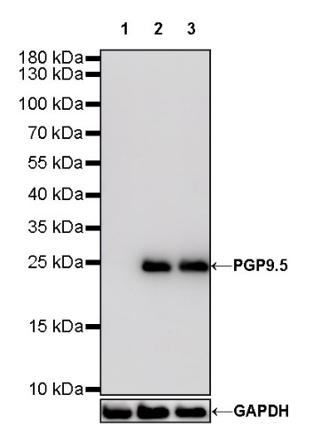
WB result of PGP9.5 Rabbit mAb
Primary antibody: PGP9.5 Rabbit mAb at 1/1000 dilution
Lane 1: K562 whole cell lysate 20 µg
Lane 2: Ramos whole cell lysate 20 µg
Lane 3: SH-SY5Y whole cell lysate 20 µg
Negative control: K562 whole cell lysate
Secondary antibody: Goat Anti-Rabbit IgG, (H+L), HRP conjugated at 1/10000 dilution
Predicted MW: 25 kDa
Observed MW: 25 kDa
WB result of PGP9.5 Rabbit mAb
Primary antibody: PGP9.5 Rabbit mAb at 1/1000 dilution
Lane 1: Neuro-2a whole cell lysate 20 µg
Secondary antibody: Goat Anti-Rabbit IgG, (H+L), HRP conjugated at 1/10000 dilution
Predicted MW: 25 kDa
Observed MW: 25 kDa
WB result of PGP9.5 Rabbit mAb
Primary antibody: PGP9.5 Rabbit mAb at 1/1000 dilution
Lane 1: C6 whole cell lysate 20 µg
Secondary antibody: Goat Anti-Rabbit IgG, (H+L), HRP conjugated at 1/10000 dilution
Predicted MW: 25 kDa
Observed MW: 25 kDa
WB result of PGP9.5 Rabbit mAb
Primary antibody : PGP9.5 Rabbit mAb at 1/1000 dilution
Lane 1 : Zebra fish lysate 20 µg
Secondary antibody: Goat Anti-Rabbit IgG, (H+L), HRP conjugated at 1/10000 dilution
Predicted MW: 25 kDa
Observed MW: 25 kDa
Flow cytometric analysis of 4% PFA fixed 90% methanol permeabilized SH-SY5Y (Human neuroblastoma epithelial cell) cells labelling PGP9.5 antibody at 1/500 (0.1 μg) dilution / (red) compared with a Rabbit monoclonal IgG (Black) isotype control and an unlabelled control (cells without incubation with primary antibody and secondary antibody) (Blue). Goat Anti - Rabbit IgG Alexa Fluor® 488 was used as the secondary antibody.
ICC shows positive staining in Neuro-2a cells. Anti-PGP9.5 antibody was used at 1/500 dilution (Green) and incubated overnight at 4°C. Goat polyclonal Antibody to Rabbit IgG - H&L (Alexa Fluor® 488) was used as secondary antibody at 1/1000 dilution. The cells were fixed with 100% ice-cold methanol and permeabilized with 0.1% PBS-Triton X-100. Nuclei were counterstained with DAPI (Blue). Counterstain with tubulin (Red).
 您现在的位置: