
PBS, 40% Glycerol, 0.05% BSA, 0.03% Proclin 300
12 months from date of receipt / reconstitution, -20 °C as supplied
| 应用 | 稀释度 | 推荐种属 |
|---|---|---|
| WB | 1:1000 | Hu, Rt |
| IP | 1:50 | Hu |
| IHC-P | 1:250 | Hu |
| ICC | 1:500 | Hu, Ms |
FAM129B, also known as MINERVA, is a protein that has been implicated in various cellular processes including cell protection, antioxidative responses, and regulation of adipogenesis. It has gained attention for its potential roles in cancer progression and as a therapeutic target. Research indicates that FAM129B can protect cardiomyocytes from hypoxia/reoxygenation-induced injury by inhibiting apoptosis, oxidative stress, and inflammatory response through the enhancement of Nrf2/ARE activation. FAM129B has been identified as an antioxidative protein that can reduce chemosensitivity by competing with Nrf2 for Keap1 binding, which influences the Nrf2/antioxidative response and is associated with poor prognosis in breast and lung cancer. FAM129B plays a crucial role during the early stages of adipocyte differentiation. It is expressed downstream of early genes such as Cebpb, Klf4, Klf5, and Srebf1a, but upstream of Pparg2, indicating its role in the regulation of adipose differentiation. FAM129B has been shown to promote tumor invasion and proliferation in non-small cell lung cancer by facilitating the phosphorylation of FAK signaling, which is associated with adverse clinical outcomes. FAM129B activates Ras, which promotes aerobic glycolysis, a process often observed in cancer cells, and this activation is important for cell cycle progression, tumor cell proliferation, and brain tumorigenesis.
WB result of FAM129B Recombinant Rabbit mAb
Primary antibody: FAM129B Recombinant Rabbit mAb at 1/1000 dilution
Lane 1: C6 whole cell lysate 20 µg
Secondary antibody: Goat Anti-rabbit IgG, (H+L), HRP conjugated at 1/10000 dilution
Predicted MW: 84 kDa
Observed MW: 100 kDa
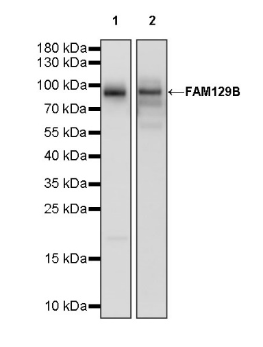
WB result of FAM129B Recombinant Rabbit mAb
Primary antibody: FAM129B Recombinant Rabbit mAb at 1/1000 dilution
Lane 1: HeLa whole cell lysate 20 µg
Lane 2: A549 whole cell lysate 20 µg
Secondary antibody: Goat Anti-rabbit IgG, (H+L), HRP conjugated at 1/10000 dilution
Predicted MW: 84 kDa
Observed MW: 95 kDa
FAM129B Rabbit mAb at 1/50 dilution (1 µg) immunoprecipitating FAM129B in 0.4 mg HeLa whole cell lysate.
Western blot was performed on the immunoprecipitate using FAM129B Rabbit mAb at 1/1000 dilution.
Secondary antibody (HRP) for IP was used at 1/1000 dilution.
Lane 1: HeLa whole cell lysate 20 µg (Input)
Lane 2: FAM129B Rabbit mAb IP in HeLa whole cell lysate
Lane 3: Rabbit monoclonal IgG IP in HeLa whole cell lysate
Predicted MW: 84 kDa
Observed MW: 95 kDa
IHC shows positive staining in paraffin-embedded human liver. Anti- FAM129B antibody was used at 1/250 dilution, followed by a HRP Polymer for Mouse & Rabbit IgG (ready to use). Counterstained with hematoxylin. Heat mediated antigen retrieval with Tris/EDTA buffer pH9.0 was performed before commencing with IHC staining protocol.
IHC shows positive staining in paraffin-embedded human placenta. Anti- FAM129B antibody was used at 1/250 dilution, followed by a HRP Polymer for Mouse & Rabbit IgG (ready to use). Counterstained with hematoxylin. Heat mediated antigen retrieval with Tris/EDTA buffer pH9.0 was performed before commencing with IHC staining protocol.
IHC shows positive staining in paraffin-embedded human hepatocellular carcinoma. Anti- FAM129B antibody was used at 1/250 dilution, followed by a HRP Polymer for Mouse & Rabbit IgG (ready to use). Counterstained with hematoxylin. Heat mediated antigen retrieval with Tris/EDTA buffer pH9.0 was performed before commencing with IHC staining protocol.
IHC shows positive staining in paraffin-embedded human lung squamous cell carcinoma. Anti- FAM129B antibody was used at 1/250 dilution, followed by a HRP Polymer for Mouse & Rabbit IgG (ready to use). Counterstained with hematoxylin. Heat mediated antigen retrieval with Tris/EDTA buffer pH9.0 was performed before commencing with IHC staining protocol.
ICC shows positive staining in HeLa cells. Anti-FAM129B antibody was used at 1/500 dilution (Green) and incubated overnight at 4°C. Goat polyclonal Antibody to Rabbit IgG - H&L (Alexa Fluor® 488) was used as secondary antibody at 1/1000 dilution. The cells were fixed with 100% ice-cold methanol and permeabilized with 0.1% PBS-Triton X-100. Nuclei were counterstained with DAPI (Blue). Counterstain with tubulin (Red).
ICC shows positive staining in NIH/3T3 cells. Anti-FAM129B antibody was used at 1/500 dilution (Green) and incubated overnight at 4°C. Goat polyclonal Antibody to Rabbit IgG - H&L (Alexa Fluor® 488) was used as secondary antibody at 1/1000 dilution. The cells were fixed with 100% ice-cold methanol and permeabilized with 0.1% PBS-Triton X-100. Nuclei were counterstained with DAPI (Blue). Counterstain with tubulin (Red).
 您现在的位置: