
12 months from date of receipt / reconstitution, -20 °C as supplied
| 应用 | 稀释度 | 推荐种属 |
|---|---|---|
| WB | 1:1000 | Hu |
| IHC-P | 1:500 | Hu |
| ICC | 1:500 | Hu |
Phosphorylation of Akt at Ser473 is a key event in the activation of the protein kinase B (PKB), commonly known as Akt. This modification is essential for the full activation of Akt and its downstream signaling pathways, which play a crucial role in regulating cell survival, growth, and metabolism. The phosphorylation at Ser473 is often used as a biomarker for the activation status of Akt. In the context of cancer, the activation of the PI3K/Akt pathway is frequently dysregulated, and Akt activity has been implicated in tumorigenesis, making it a potential therapeutic target. The level of phosphorylated Akt (Ser473) has been correlated with clinical outcomes in various cancers, although the specific associations can vary between different tumor types and studies. Moreover, the phosphorylation of Akt at Ser473 is regulated by various cellular stresses, including endoplasmic reticulum (ER) stress. ER stress can modulate Akt substrate specificity and activity in a severity-dependent manner, which has implications for the development of therapeutic strategies targeting the Akt pathway.
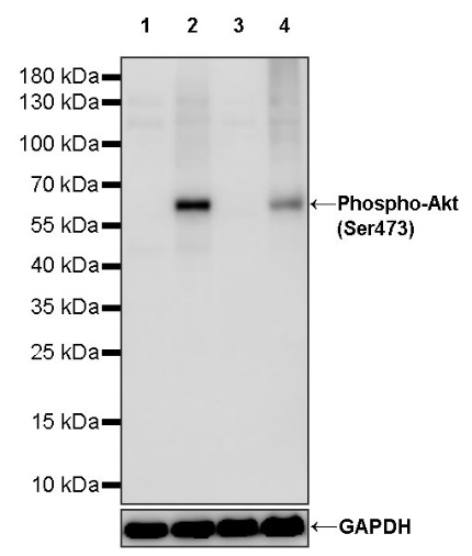
WB result of Phospho-Akt (Ser473) Recombinant Rabbit mAb
Primary antibody: Phospho-Akt (Ser473) Recombinant Rabbit mAb at 1/1000 dilution
Lane 1: untreated Jurkat whole cell lysate 20 µg
Lane 2: Jurkat treated with 100 nM Calyculin A for 30 minutes whole cell lysate 20 µg
Lane 3: untreated HeLa whole cell lysate 20 µg
Lane 4: HeLa treated with 100 ng/ml Calyculin A for 30 minutes whole cell lysate 20 µg
Secondary antibody: Goat Anti-rabbit IgG, (H+L), HRP conjugated at 1/10000 dilution
Predicted MW: 56 kDa
Observed MW: 60 kDa
IHC shows positive staining in paraffin-embedded human esophageal cancer and negative staining in esophageal cancer treated with phosphatase. Anti-Phospho-Akt (Ser473) antibody was used at 1/500 dilution, followed by a HRP Polymer for Mouse & Rabbit IgG (ready to use). Counterstained with hematoxylin. Heat mediated antigen retrieval with Tris/EDTA buffer pH9.0 was performed before commencing with IHC staining protocol.
IHC shows positive staining in paraffin-embedded human endometrial cancer and negative staining in esophageal cancer treated with phosphatase. Anti-Phospho-Akt (Ser473) antibody was used at 1/500 dilution, followed by a HRP Polymer for Mouse & Rabbit IgG (ready to use). Counterstained with hematoxylin. Heat mediated antigen retrieval with Tris/EDTA buffer pH9.0 was performed before commencing with IHC staining protocol.
ICC analysis of Jurkat cells treated with Calyculin A (100nM, 30 min) (top panel) and Jurkat cells untreated with Calyculin A (100nM, 30 min) (below panel). Anti-Phospho-Akt (Ser473) antibody was used at 1/500 dilution (Green) and incubated overnight at 4°C. Goat polyclonal Antibody to Rabbit IgG - H&L (Alexa Fluor® 488) was used as secondary antibody at 1/1000 dilution. The cells were fixed with 4% PFA and permeabilized with 0.1% PBS-Triton X-100. Nuclei were counterstained with DAPI (Blue). Counterstain with tubulin (Red).
 您现在的位置: