
PBS, 40% Glycerol, 0.05% BSA, 0.03% Proclin 300
12 months from date of receipt / reconstitution, -20 °C as supplied
| 应用 | 稀释度 | 推荐种属 |
|---|---|---|
| WB | 1:1000 | Hu, Ms, Rt |
| IP | 1:50 | Hu |
| IHC-P | 1:1000 | Hu, Ms, Rt |
| ICC | 1:500 | Hu, Ms |
YTHDF2 is an RNA binding protein that acts as a reader of N6-methyladenosine (m6A) modifications on RNA molecules. The protein has been implicated in various cellular processes, including RNA metabolism, and it has been linked to the progression of several types of cancer. Research has shown that YTHDF2 can promote cancer progression by increasing its binding affinity with m6A-modified mRNAs, leading to mRNA degradation. It has also been reported that YTHDF2 can be post-translationally modified, such as through SUMOylation, which can enhance its function and contribute to tumor growth. Furthermore, YTHDF2 has been identified as a potential therapeutic target in cancer treatment. Its expression or functional abnormalities are associated with the occurrence and metastasis of various tumors, suggesting its role as a potential oncogene and a therapeutic target. In addition to its role in cancer, YTHDF2 has been shown to modulate the role of M2 macrophages in the origin of metastatic tumors and to control antitumor immunity through CD8+ T cells.
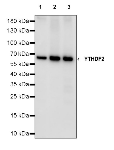
WB result of YTHDF2 Recombinant Rabbit mAb
Primary antibody: YTHDF2 Recombinant Rabbit mAb at 1/1000 dilution
Lane 1: HEK-293 whole cell lysate 20 µg
Lane 2: HeLa whole cell lysate 20 µg
Lane 3: HCT 116 whole cell lysate 20 µg
Secondary antibody: Goat Anti-rabbit IgG, (H+L), HRP conjugated at 1/10000 dilution
Predicted MW: 62 kDa
Observed MW: 62 kDa
WB result of YTHDF2 Recombinant Rabbit mAb
Primary antibody: YTHDF2 Recombinant Rabbit mAb at 1/1000 dilution
Lane 1: NIH/3T3 whole cell lysate 20 µg
Lane 2: RAW264.7 whole cell lysate 20 µg
Secondary antibody: Goat Anti-rabbit IgG, (H+L), HRP conjugated at 1/10000 dilution
Predicted MW: 62 kDa
Observed MW: 62 kDa
WB result of YTHDF2 Recombinant Rabbit mAb
Primary antibody: YTHDF2 Recombinant Rabbit mAb at 1/1000 dilution
Lane 1: PC-12 whole cell lysate 20 µg
Lane 2: rat testis lysate 20 µg
Secondary antibody: Goat Anti-rabbit IgG, (H+L), HRP conjugated at 1/10000 dilution
Predicted MW: 62 kDa
Observed MW: 62 kDa
Flow cytometric analysis of 4% PFA fixed 90% methanol permeabilized HeLa (Human cervix adenocarcinoma epithelial cell) labelling YTHDF2 antibody at 1/500 dilution (0.1 μg) / (Red) compared with a Rabbit monoclonal IgG (Black) isotype control and an unlabelled control (cells without incubation with primary antibody and secondary antibody) (Blue). Goat Anti - Rabbit IgG Alexa Fluor® 488 was used as the secondary antibody.
Flow cytometric analysis of 4% PFA fixed 90% methanol permeabilized NIH/3T3 (Mouse embryonic fibroblast) labelling YTHDF2 antibody at 1/500 dilution (0.1 μg) / (Red) compared with a Rabbit monoclonal IgG (Black) isotype control and an unlabelled control (cells without incubation with primary antibody and secondary antibody) (Blue). Goat Anti - Rabbit IgG Alexa Fluor® 488 was used as the secondary antibody.
YTHDF2 Rabbit mAb at 1/50 dilution (1 µg) immunoprecipitating YTHDF2 in 0.4 mg HeLa whole cell lysate.
Western blot was performed on the immunoprecipitate using YTHDF2 Rabbit mAb at 1/1000 dilution.
Secondary antibody (HRP) for IP was used at 1/1000 dilution.
Lane 1: HeLa whole cell lysate 20 µg (Input)
Lane 2: YTHDF2 Rabbit mAb IP in HeLa whole cell lysate
Lane 3: Rabbit monoclonal IgG IP in HeLa whole cell lysate
Predicted MW: 62 kDa
Observed MW: 62 kDa
IHC shows positive staining in paraffin-embedded human testis. Anti- YTHDF2 antibody was used at 1/1000 dilution, followed by a HRP Polymer for Mouse & Rabbit IgG (ready to use). Counterstained with hematoxylin. Heat mediated antigen retrieval with Tris/EDTA buffer pH9.0 was performed before commencing with IHC staining protocol.
IHC shows positive staining in paraffin-embedded mouse cerebral cortex. Anti- YTHDF2 antibody was used at 1/1000 dilution, followed by a HRP Polymer for Mouse & Rabbit IgG (ready to use). Counterstained with hematoxylin. Heat mediated antigen retrieval with Tris/EDTA buffer pH9.0 was performed before commencing with IHC staining protocol.
IHC shows positive staining in paraffin-embedded mouse testis. Anti- YTHDF2 antibody was used at 1/1000 dilution, followed by a HRP Polymer for Mouse & Rabbit IgG (ready to use). Counterstained with hematoxylin. Heat mediated antigen retrieval with Tris/EDTA buffer pH9.0 was performed before commencing with IHC staining protocol.
IHC shows positive staining in paraffin-embedded rat cerebral cortex. Anti- YTHDF2 antibody was used at 1/1000 dilution, followed by a HRP Polymer for Mouse & Rabbit IgG (ready to use). Counterstained with hematoxylin. Heat mediated antigen retrieval with Tris/EDTA buffer pH9.0 was performed before commencing with IHC staining protocol.
IHC shows positive staining in paraffin-embedded rat testis. Anti- YTHDF2 antibody was used at 1/1000 dilution, followed by a HRP Polymer for Mouse & Rabbit IgG (ready to use). Counterstained with hematoxylin. Heat mediated antigen retrieval with Tris/EDTA buffer pH9.0 was performed before commencing with IHC staining protocol.
ICC shows positive staining in HeLa cells. Anti-YTHDF2 antibody was used at 1/500 dilution (Green) and incubated overnight at 4°C. Goat polyclonal Antibody to Rabbit IgG - H&L (Alexa Fluor® 488) was used as secondary antibody at 1/1000 dilution. The cells were fixed with 4% PFA and permeabilized with 0.1% PBS-Triton X-100. Nuclei were counterstained with DAPI (Blue). Counterstain with tubulin (Red).
ICC shows positive staining in NIH/3T3 cells. Anti-YTHDF2 antibody was used at 1/500 dilution (Green) and incubated overnight at 4°C. Goat polyclonal Antibody to Rabbit IgG - H&L (Alexa Fluor® 488) was used as secondary antibody at 1/1000 dilution. The cells were fixed with 4% PFA and permeabilized with 0.1% PBS-Triton X-100. Nuclei were counterstained with DAPI (Blue). Counterstain with tubulin (Red).
Expression of YTHDF2 in tumor tissue
 您现在的位置: