
12 months from date of receipt / reconstitution, -20 °C as supplied
| 应用 | 稀释度 | 推荐种属 |
|---|---|---|
| WB | 1:4000 | Hu, Ms, Rt |
| IHC-P | 1:250 | Hu, Ms, Rt |
| ICC | 1:2000 | Hu |
| ICFCM | 1:2000 | Hu |
ATG9A is an autophagy-related protein that serves as a multi-spanning membrane protein, playing a crucial role in the process of autophagy. It aids in the expansion of phagophore membranes during the early stages of autophagy by flipping phospholipids between the two membrane leaflets, which assists in the formation of autophagosomes. Research has indicated that the absence of ATG9A not only inhibits autophagy but also increases the size and number of lipid droplets in human cell lines. Furthermore, the depletion of ATG9A prevents the transfer of fatty acids from lipid droplets to mitochondria, thereby hindering the utilization of fatty acids in mitochondrial respiration. ATG9A also plays a role in intracellular lipid homeostasis, particularly in relation to lipid droplets. The knockout or knockdown of ATG9A leads to an increase in the number and size of lipid droplets, and impedes the transport of fatty acids from lipid droplets to mitochondria, resulting in decreased mitochondrial fatty acid beta-oxidation. Additionally, ATG9A is closely associated with a subdomain of the endoplasmic reticulum that is rich in TMEM41B, and both structures are found near phagophores, lipid droplets, and mitochondria, highlighting the key role of ATG9A in lipid mobilization. Under heat stress conditions, ATG9A is involved in the regulation of Golgi apparatus dynamics and its functional molecular mechanisms.
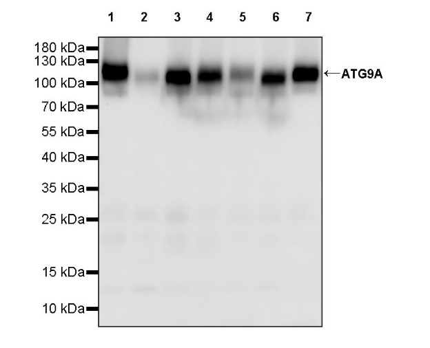
WB result of ATG9A Recombinant Rabbit mAb
Primary antibody: ATG9A Recombinant Rabbit mAb at 1/4000 dilution
Lane 1: unboiled HepG2 whole cell lysate 20 µg
Lane 2: unboiled MCF7 whole cell lysate 20 µg
Lane 3: unboiled 293T whole cell lysate 20 µg
Lane 4: unboiled A375 whole cell lysate 20 µg
Lane 5: unboiled DU 145 whole cell lysate 20 µg
Lane 6: unboiled HEK-293 whole cell lysate 20 µg
Lane 7: unboiled HeLa whole cell lysate 20 µg
Secondary antibody: Goat Anti-rabbit IgG, (H+L), HRP conjugated at 1/10000 dilution
Predicted MW: 94 kDa
Observed MW: 100~130 kDa
WB result of ATG9A Recombinant Rabbit mAb
Primary antibody: ATG9A Recombinant Rabbit mAb at 1/4000 dilution
Lane 1: unboiled Neuro-2a whole cell lysate 20 µg
Lane 2: unboiled mouse brain lysate 20 µg
Secondary antibody: Goat Anti-rabbit IgG, (H+L), HRP conjugated at 1/10000 dilution
Predicted MW: 94 kDa
Observed MW: 95~120 kDa
WB result of ATG9A Recombinant Rabbit mAb
Primary antibody: ATG9A Recombinant Rabbit mAb at 1/4000 dilution
Lane 1: unboiled rat brain lysate 20 µg
Secondary antibody: Goat Anti-rabbit IgG, (H+L), HRP conjugated at 1/10000 dilution
Predicted MW: 94 kDa
Observed MW: 90~120 kDa
Flow cytometric analysis of 4% PFA fixed 90% methanol permeabilized HepG2 (Human hepatocellular carcinoma epithelial cell) labelling ATG9A antibody at 1/2000 dilution (0.1 μg) / (Red) compared with a Rabbit monoclonal IgG (Black) isotype control and an unlabelled control (cells without incubation with primary antibody and secondary antibody) (Blue). Goat Anti - Rabbit IgG Alexa Fluor® 488 was used as the secondary antibody.
IHC shows positive staining in paraffin-embedded human testis. Anti-ATG9A antibody was used at 1/250 dilution, followed by a HRP Polymer for Mouse & Rabbit IgG (ready to use). Counterstained with hematoxylin. Heat mediated antigen retrieval with Tris/EDTA buffer pH9.0 was performed before commencing with IHC staining protocol.
IHC shows positive staining in paraffin-embedded human hepatocellular carcinoma. Anti-ATG9A antibody was used at 1/250 dilution, followed by a HRP Polymer for Mouse & Rabbit IgG (ready to use). Counterstained with hematoxylin. Heat mediated antigen retrieval with Tris/EDTA buffer pH9.0 was performed before commencing with IHC staining protocol.
IHC shows positive staining in paraffin-embedded human cervical squamous cell carcinoma. Anti-ATG9A antibody was used at 1/250 dilution, followed by a HRP Polymer for Mouse & Rabbit IgG (ready to use). Counterstained with hematoxylin. Heat mediated antigen retrieval with Tris/EDTA buffer pH9.0 was performed before commencing with IHC staining protocol.
IHC shows positive staining in paraffin-embedded human endometrial carcinoma. Anti-ATG9A antibody was used at 1/250 dilution, followed by a HRP Polymer for Mouse & Rabbit IgG (ready to use). Counterstained with hematoxylin. Heat mediated antigen retrieval with Tris/EDTA buffer pH9.0 was performed before commencing with IHC staining protocol.
IHC shows positive staining in paraffin-embedded human lung squamous cell carcinoma. Anti-ATG9A antibody was used at 1/250 dilution, followed by a HRP Polymer for Mouse & Rabbit IgG (ready to use). Counterstained with hematoxylin. Heat mediated antigen retrieval with Tris/EDTA buffer pH9.0 was performed before commencing with IHC staining protocol.
IHC shows positive staining in paraffin-embedded mouse kidney. Anti-ATG9A antibody was used at 1/250 dilution, followed by a HRP Polymer for Mouse & Rabbit IgG (ready to use). Counterstained with hematoxylin. Heat mediated antigen retrieval with Tris/EDTA buffer pH9.0 was performed before commencing with IHC staining protocol.
IHC shows positive staining in paraffin-embedded rat cerebral cortex. Anti-ATG9A antibody was used at 1/250 dilution, followed by a HRP Polymer for Mouse & Rabbit IgG (ready to use). Counterstained with hematoxylin. Heat mediated antigen retrieval with Tris/EDTA buffer pH9.0 was performed before commencing with IHC staining protocol.
ICC shows positive staining in HepG2 cells. Anti-ATG9A antibody was used at 1/2000 dilution (Green) and incubated overnight at 4°C. Goat polyclonal Antibody to Rabbit IgG - H&L (Alexa Fluor® 488) was used as secondary antibody at 1/1000 dilution. The cells were fixed with 100% ice-cold methanol and permeabilized with 0.1% PBS-Triton X-100. Nuclei were counterstained with DAPI (Blue). Counterstain with tubulin (Red).
Expression of ATG9A in tumor tissue
Expression of ATG9A in human tissue
Expression of ATG9A in mouse & rat tissue
 您现在的位置: