
12 months from date of receipt / reconstitution, -20 °C as supplied
| 应用 | 稀释度 | 推荐种属 |
|---|---|---|
| WB | 1:50000 | Hu, Ms, Rt |
| IHC-P | 1:1000 | Hu, Ms, Rt |
| ICC | 1:4000 | Hu, Ms |
Beta IV Tubulin, also known as tubulin beta 4A class IVa or TUBB4, is a specific isotype of beta-tubulin that plays a crucial role in the central nervous system. It is selectively expressed by oligodendrocytes, which are the cells responsible for myelination in the brain and spinal cord. This tubulin isotype is important for maintaining the structure and function of oligodendrocytes and their ability to form myelin. Myelin is a fatty substance that insulates nerve fibers and is essential for the proper conduction of nerve impulses. Beta IV Tubulin may also be involved in cell signaling pathways and gene expression patterns that change during oligodendrocyte differentiation and myelination.
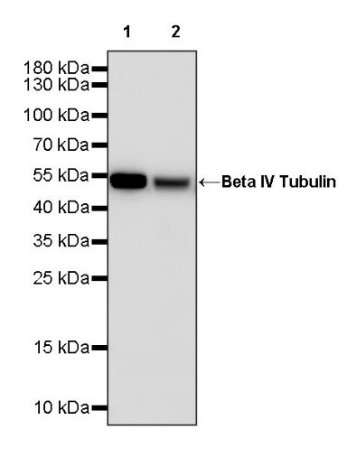
WB result of Beta IV Tubulin Rabbit pAb
Primary antibody: Beta IV Tubulin Rabbit pAb at 1/50000 dilution
Lane 1: HeLa whole cell lysate 20 µg
Lane 2: Jurkat whole cell lysate 20 µg
Secondary antibody: Goat Anti-rabbit IgG, (H+L), HRP conjugated at 1/10000 dilution
Predicted MW: 50 kDa
Observed MW: 52 kDa
WB result of Beta IV Tubulin Rabbit pAb
Primary antibody: Beta IV Tubulin Rabbit pAb at 1/50000 dilution
Lane 1: Neuro-2a whole cell lysate 20 µg
Lane 2: mouse brain lysate 20 µg
Secondary antibody: Goat Anti-rabbit IgG, (H+L), HRP conjugated at 1/10000 dilution
Predicted MW: 50 kDa
Observed MW: 52 kDa
WB result of Beta IV Tubulin Rabbit pAb
Primary antibody: Beta IV Tubulin Rabbit pAb at 1/50000 dilution
Lane 1: C6 whole cell lysate 20 µg
Lane 2: rat brain lysate 20 µg
Secondary antibody: Goat Anti-rabbit IgG, (H+L), HRP conjugated at 1/10000 dilution
Predicted MW: 50 kDa
Observed MW: 52 kDa
IHC shows positive staining in paraffin-embedded human tonsil. Anti- Beta IV Tubulin antibody was used at 1/1000 dilution, followed by a HRP Polymer for Mouse & Rabbit IgG (ready to use). Counterstained with hematoxylin. Heat mediated antigen retrieval with Tris/EDTA buffer pH9.0 was performed before commencing with IHC staining protocol.
IHC shows positive staining in paraffin-embedded human kidney. Anti- Beta IV Tubulin antibody was used at 1/1000 dilution, followed by a HRP Polymer for Mouse & Rabbit IgG (ready to use). Counterstained with hematoxylin. Heat mediated antigen retrieval with Tris/EDTA buffer pH9.0 was performed before commencing with IHC staining protocol.
IHC shows positive staining in paraffin-embedded human colon cancer. Anti- Beta IV Tubulin antibody was used at 1/1000 dilution, followed by a HRP Polymer for Mouse & Rabbit IgG (ready to use). Counterstained with hematoxylin. Heat mediated antigen retrieval with Tris/EDTA buffer pH9.0 was performed before commencing with IHC staining protocol.
IHC shows positive staining in paraffin-embedded mouse kidney. Anti- Beta IV Tubulin antibody was used at 1/1000 dilution, followed by a HRP Polymer for Mouse & Rabbit IgG (ready to use). Counterstained with hematoxylin. Heat mediated antigen retrieval with Tris/EDTA buffer pH9.0 was performed before commencing with IHC staining protocol.
IHC shows positive staining in paraffin-embedded rat cerebral cortex. Anti- Beta IV Tubulin antibody was used at 1/1000 dilution, followed by a HRP Polymer for Mouse & Rabbit IgG (ready to use). Counterstained with hematoxylin. Heat mediated antigen retrieval with Tris/EDTA buffer pH9.0 was performed before commencing with IHC staining protocol.
ICC shows positive staining in HeLa cells. Anti-Beta IV Tubulin antibody was used at 1/4000 dilution (Green) and incubated overnight at 4°C. Goat polyclonal Antibody to Rabbit IgG - H&L (Alexa Fluor® 488) was used as secondary antibody at 1/1000 dilution. The cells were fixed with 4% PFA and permeabilized with 0.1% PBS-Triton X-100. Nuclei were counterstained with DAPI (Blue).
ICC shows positive staining in Neuro-2a cells. Anti-Beta IV Tubulin antibody was used at 1/4000 dilution (Green) and incubated overnight at 4°C. Goat polyclonal Antibody to Rabbit IgG - H&L (Alexa Fluor® 488) was used as secondary antibody at 1/1000 dilution. The cells were fixed with 4% PFA and permeabilized with 0.1% PBS-Triton X-100. Nuclei were counterstained with DAPI (Blue).
Expression of Beta IV Tubulin in tumor tissue
Expression of Beta IV Tubulin in human tissue
Expression of Beta IV Tubulin in mouse & rat tissue
 您现在的位置: