
PBS, 40% Glycerol, 0.05% BSA, 0.03% Proclin 300
12 months from date of receipt / reconstitution, -20 °C as supplied
| 应用 | 稀释度 | 推荐种属 |
|---|---|---|
| WB | 1:1000 | Hu |
| IP | 1:50 | Hu |
| IHC-P | 1:500 | Hu |
Intercellular adhesion molecule 3 (ICAM3) also known as CD50 (Cluster of Differentiation 50), is a protein that in humans is encoded by the ICAM3 gene. The protein is constitutively expressed on the surface of leukocytes, which are also called white blood cells and are part of the immune system. ICAM3 mediates adhesion between cells by binding to specific integrin receptors. It plays an important role in the immune cell response through its facilitation of interactions between T cells and dendritic cells, which allows for T cell activation. ICAM3 also mediates the clearance of cells undergoing apoptosis by attracting and binding macrophages, a type of cell that breaks down infected or dying cells through a process known as phagocytosis, to apoptotic cells.
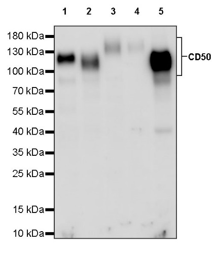
WB result of CD50 Rabbit pAb
Primary antibody: CD50 Rabbit pAb at 1/1000 dilution
Lane 1: Jurkat whole cell lysate 20 µg
Lane 2: Ramos whole cell lysate 20 µg
Lane 3: THP-1 whole cell lysate 20 µg
Lane 4: HL-60 whole cell lysate 20 µg
Lane 5: HuT 78 whole cell lysate 20 µg
Secondary antibody: Goat Anti-rabbit IgG, (H+L), HRP conjugated at 1/10000 dilution
Predicted MW: 60 kDa
Observed MW: 100~160 kDa
Flow cytometric analysis of human PBMC (human peripheral blood mononuclear cell) labelling Human CD50 antibody at 1/50 (1 μg) dilution (Right) compared with a Rabbit monoclonal IgG isotype control (Left). Goat Anti - Rabbit IgG Alexa Fluor® 488 was used as the secondary antibody. Then cells were stained with CD3 - Brilliant Violet 421™ separately. Gated on total viable cells.
CD50 Rabbit pAb at 1/50 dilution (1 µg) immunoprecipitating CD50 in 0.4 mg Jurkat whole cell lysate.
Western blot was performed on the immunoprecipitate using CD50 Rabbit pAb at 1/1000 dilution.
Secondary antibody (HRP) for IP was used at 1/1000 dilution.
Lane 1: Jurkat whole cell lysate 20 µg (Input)
Lane 2: CD50 Rabbit pAb IP in Jurkat whole cell lysate
Lane 3: Rabbit monoclonal IgG IP in Jurkat whole cell lysate
Predicted MW: 60 kDa
Observed MW: 100~160 kDa
IHC shows positive staining in paraffin-embedded human spleen. Anti-CD50 antibody was used at 1/500 dilution, followed by a HRP Polymer for Mouse & Rabbit IgG (ready to use). Counterstained with hematoxylin. Heat mediated antigen retrieval with Tris/EDTA buffer pH9.0 was performed before commencing with IHC staining protocol.
IHC shows positive staining in paraffin-embedded human stomach. Anti-CD50 antibody was used at 1/500 dilution, followed by a HRP Polymer for Mouse & Rabbit IgG (ready to use). Counterstained with hematoxylin. Heat mediated antigen retrieval with Tris/EDTA buffer pH9.0 was performed before commencing with IHC staining protocol.
IHC shows positive staining in paraffin-embedded human tonsil. Anti-CD50 antibody was used at 1/500 dilution, followed by a HRP Polymer for Mouse & Rabbit IgG (ready to use). Counterstained with hematoxylin. Heat mediated antigen retrieval with Tris/EDTA buffer pH9.0 was performed before commencing with IHC staining protocol.
IHC shows positive staining in paraffin-embedded human colon cancer. Anti-CD50 antibody was used at 1/500 dilution, followed by a HRP Polymer for Mouse & Rabbit IgG (ready to use). Counterstained with hematoxylin. Heat mediated antigen retrieval with Tris/EDTA buffer pH9.0 was performed before commencing with IHC staining protocol.
IHC shows positive staining in paraffin-embedded human lung squamous cell carcinoma. Anti-CD50 antibody was used at 1/500 dilution, followed by a HRP Polymer for Mouse & Rabbit IgG (ready to use). Counterstained with hematoxylin. Heat mediated antigen retrieval with Tris/EDTA buffer pH9.0 was performed before commencing with IHC staining protocol.
IHC shows positive staining in paraffin-embedded human ovarian carcinoma. Anti-CD50 antibody was used at 1/500 dilution, followed by a HRP Polymer for Mouse & Rabbit IgG (ready to use). Counterstained with hematoxylin. Heat mediated antigen retrieval with Tris/EDTA buffer pH9.0 was performed before commencing with IHC staining protocol.
Expression of CD50 in tumor tissue
Expression of CD50 in human tissue
Expression of CD50 in mouse & rat tissue
 您现在的位置: