
PBS, 40% Glycerol, 0.05% BSA, 0.03% Proclin 300
12 months from date of receipt / reconstitution, -20 °C as supplied
| 应用 | 稀释度 | 推荐种属 |
|---|---|---|
| WB | 1:1000 | Hu |
| IP | 1:50 | Hu |
| IHC-P | 1:250 | Hu, Ms, Rt |
| ICC | 1:500 | Hu |
c-Fos is a nuclear phosphoprotein that plays a pivotal role in cell signaling, proliferation, and differentiation. It forms a heterodimer with c-Jun, which then forms the activator protein-1 (AP-1) complex. This complex binds to specific DNA sequences in the promoter and enhancer regions of target genes, transforming extracellular signals into changes in gene expression. c-Fos is crucial in regulating cell development and maintaining skeletal formation. The expression of c-Fos is associated with various biological processes, including cell proliferation, differentiation, and programmed cell death. Additionally, c-Fos is involved in learning and memory processes, with changes in its expression observed in brain regions associated with recognition, working, and fear-related memory. c-Fos is also used as a marker for neural activity, with its expression linked to changes in neuronal function and plasticity. The expression of c-Fos is also related to the occurrence and development of cancer. The c-Fos protein can serve as a marker for tumorigenesis, and its dysregulation is associated with cancer progression. The absence of c-Fos leads to significant neurological abnormalities in mice embryos during development, indicating that c-Fos plays an important role in neurogenesis and brain development.
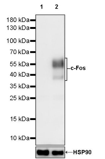
WB result of c-Fos Recombinant Rabbit mAb
Primary antibody: c-Fos Recombinant Rabbit mAb at 1/1000 dilution
Lane 1: untreated HeLa whole cell lysate 20 µg
Lane 2: HeLa starve overnight, then treated with 200 nM PMA for 4 hours whole cell lysate 20 µg
Secondary antibody: Goat Anti-rabbit IgG, (H+L), HRP conjugated at 1/10000 dilution
Predicted MW: 41 kDa
Observed MW: 38~60 kDa
c-Fos Recombinant Rabbit mAb at 1/50 dilution (1 µg) immunoprecipitating c-Fos in 0.4 mg HeLa starve overnight, 200nM PMA for 4 hours whole cell lysate whole cell lysate.
Western blot was performed on the immunoprecipitate using c-Fos Recombinant Rabbit mAb at 1/1000 dilution.
Secondary antibody (HRP) for IP was used at 1/1000 dilution.
Lane 1: HeLa starve overnight, 200nM PMA for 4 hours whole cell lysate whole cell lysate 40 µg (Input)
Lane 2: c-Fos Recombinant Rabbit mAb IP in HeLa starve overnight, 200nM PMA for 4 hours whole cell lysate whole cell lysate
Lane 3: Rabbit monoclonal IgG IP in HeLa starve overnight, 200nM PMA for 4 hours whole cell lysate whole cell lysate
Predicted MW: 41 kDa
Observed MW: 38~60 kDa
Exposure time: 20 s
IHC shows positive staining in paraffin-embedded human colon. Anti- c-Fos antibody was used at 1/250 dilution, followed by a HRP Polymer for Mouse & Rabbit IgG (ready to use). Counterstained with hematoxylin. Heat mediated antigen retrieval with Tris/EDTA buffer pH9.0 was performed before commencing with IHC staining protocol.
IHC shows positive staining in paraffin-embedded human stomach. Anti- c-Fos antibody was used at 1/250 dilution, followed by a HRP Polymer for Mouse & Rabbit IgG (ready to use). Counterstained with hematoxylin. Heat mediated antigen retrieval with Tris/EDTA buffer pH9.0 was performed before commencing with IHC staining protocol.
IHC shows positive staining in paraffin-embedded human lung adenocarcinoma. Anti- c-Fos antibody was used at 1/250 dilution, followed by a HRP Polymer for Mouse & Rabbit IgG (ready to use). Counterstained with hematoxylin. Heat mediated antigen retrieval with Tris/EDTA buffer pH9.0 was performed before commencing with IHC staining protocol.
IHC shows positive staining in paraffin-embedded mouse stomach. Anti- c-Fos antibody was used at 1/250 dilution, followed by a HRP Polymer for Mouse & Rabbit IgG (ready to use). Counterstained with hematoxylin. Heat mediated antigen retrieval with Tris/EDTA buffer pH9.0 was performed before commencing with IHC staining protocol.
IHC shows positive staining in paraffin-embedded rat colon. Anti- c-Fos antibody was used at 1/250 dilution, followed by a HRP Polymer for Mouse & Rabbit IgG (ready to use). Counterstained with hematoxylin. Heat mediated antigen retrieval with Tris/EDTA buffer pH9.0 was performed before commencing with IHC staining protocol.
ICC analysis of HeLa cells starve overnight then treated with 200nM PMA for 4 hours whole cell lysate and untreated HeLa cells (below panel). Anti- c-Fos antibody was used at 1/500 dilution (Green) and incubated overnight at 4°C. Goat polyclonal Antibody to Rabbit IgG - H&L (Alexa Fluor® 488) was used as secondary antibody at 1/1000 dilution. The cells were fixed with 4% PFA and permeabilized with 0.1% PBS-Triton X-100. Nuclei were counterstained with DAPI (Blue). Counterstain with tubulin (Red).
Expression of c-Fos in tumor tissue
 您现在的位置: