
12 months from date of receipt / reconstitution, -20 °C as supplied
| 应用 | 稀释度 |
|---|---|
| WB | 1:1000 |
| IP | 1:50 |
| IHC-P | 1:250 |
| ICC | 1:500 |
LEF1 (Lymphoid Enhancer-Binding Factor 1) is a transcription factor belonging to the High-Mobility Group (HMG) family of proteins, which plays a role in various biological processes, including cell proliferation, differentiation, and survival. LEF1 is a key downstream effector of the Wnt/β-catenin signaling pathway and can regulate the transcription of target genes by binding with β-catenin. In cancer research, the abnormal expression of LEF1 is associated with the occurrence and progression of various types of cancer. It also plays a role in stem cell maintenance and organ development, particularly in the process of Epithelial-Mesenchymal Transition (EMT), where it activates the transcription of EMT effectors such as N-Cadherin, Vimentin, and Snail. In certain cancer cell types, such as Chronic Lymphocytic Leukemia (CLL), Burkitt's Lymphoma (BL), Acute Lymphoblastic Leukemia (ALL), Oral Squamous Cell Carcinoma (OSCC), and Colorectal Cancer (CRC), the activity of LEF1 makes it a valuable biomarker for predicting patient prognosis. Furthermore, LEF1 promotes the expression and activity of the androgen receptor in prostate cancer in an androgen-independent manner, ultimately increasing the growth of prostate cancer regardless of androgen ablation therapy. LEF1's inhibition or knockdown has been shown to slow down cancer growth, migration, and invasion, making it a potential target for cancer treatment. In the context of hematological malignancies, the expression and function of LEF1 differ between normal and leukemic hematopoiesis, and its expression in Chronic Lymphocytic Leukemia (CLL) is closely related to the progression and prognosis of the disease. The upregulation of LEF1 in CLL/Small Lymphocytic Lymphoma (SLL) may begin at an early stage of the disease.
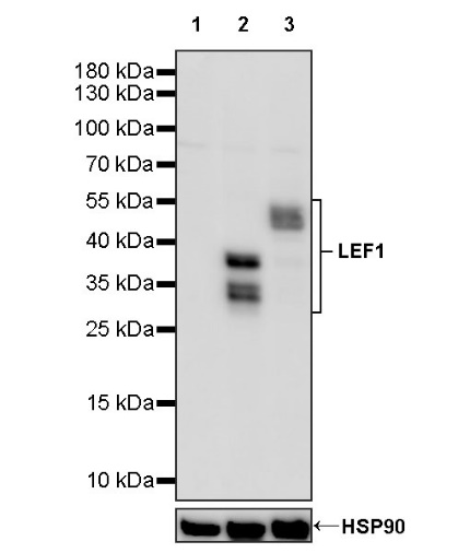
WB result of LEF1 Recombinant Rabbit mAb
Primary antibody: LEF1 Recombinant Rabbit mAb at 1/1000 dilution
Lane 1: LNCaP whole cell lysate 20 µg
Lane 2: Jurkat whole cell lysate 20 µg
Lane 3: Ramos whole cell lysate 20 µg
Negative control: LNCaP whole cell lysate
Secondary antibody: Goat Anti-rabbit IgG, (H+L), HRP conjugated at 1/10000 dilution
Predicted MW: 44 kDa
Observed MW: 30~55 kDa
WB result of LEF1 Recombinant Rabbit mAb
Primary antibody: LEF1 Recombinant Rabbit mAb at 1/1000 dilution
Lane 1: mouse thymus lysate 20 µg
Secondary antibody: Goat Anti-rabbit IgG, (H+L), HRP conjugated at 1/10000 dilution
Predicted MW: 44 kDa
Observed MW: 30~55 kDa
WB result of LEF1 Recombinant Rabbit mAb
Primary antibody: LEF1 Recombinant Rabbit mAb at 1/1000 dilution
Lane 1: rat thymus lysate 20 µg
Secondary antibody: Goat Anti-rabbit IgG, (H+L), HRP conjugated at 1/10000 dilution
Predicted MW: 44 kDa
Observed MW: 30~55 kDa
LEF1 Rabbit mAb at 1/50 dilution (1 µg) immunoprecipitating LEF1 in 0.4 mg Jurkat whole cell lysate.
Western blot was performed on the immunoprecipitate using LEF1 Rabbit mAb at 1/1000 dilution.
Secondary antibody (HRP) for IP was used at 1/1000 dilution.
Lane 1: Jurkat whole cell lysate 20 µg (Input)
Lane 2: LEF1 Rabbit mAb IP in Jurkat whole cell lysate
Lane 3: Rabbit monoclonal IgG IP in Jurkat whole cell lysate
Predicted MW: 44 kDa
Observed MW: 30~55 kDa
IHC shows positive staining in paraffin-embedded human thymus. Anti-LEF1 antibody was used at 1/250 dilution, followed by a HRP Polymer for Mouse & Rabbit IgG (ready to use). Counterstained with hematoxylin. Heat mediated antigen retrieval with Tris/EDTA buffer pH9.0 was performed before commencing with IHC staining protocol.
IHC shows positive staining in paraffin-embedded human thymus. Anti-LEF1 antibody was used at 1/250 dilution, followed by a HRP Polymer for Mouse & Rabbit IgG (ready to use). Counterstained with hematoxylin. Heat mediated antigen retrieval with Tris/EDTA buffer pH9.0 was performed before commencing with IHC staining protocol.
IHC shows positive staining in paraffin-embedded mouse spleen. Anti-LEF1 antibody was used at 1/250 dilution, followed by a HRP Polymer for Mouse & Rabbit IgG (ready to use). Counterstained with hematoxylin. Heat mediated antigen retrieval with Tris/EDTA buffer pH9.0 was performed before commencing with IHC staining protocol.
IHC shows positive staining in paraffin-embedded rat spleen. Anti-LEF1 antibody was used at 1/250 dilution, followed by a HRP Polymer for Mouse & Rabbit IgG (ready to use). Counterstained with hematoxylin. Heat mediated antigen retrieval with Tris/EDTA buffer pH9.0 was performed before commencing with IHC staining protocol.
ICC shows positive staining in Jurkat cells (top panel) and negative staining in LNcap cells (below panel). Anti-LEF1 antibody was used at 1/500 dilution (Green) and incubated overnight at 4°C. Goat polyclonal Antibody to Rabbit IgG - H&L (Alexa Fluor® 488) was used as secondary antibody at 1/1000 dilution. The cells were fixed with 4% PFA and permeabilized with 0.1% PBS-Triton X-100. Nuclei were counterstained with DAPI (Blue). Counterstain with tubulin (Red).
 您现在的位置: