
PBS, 40% Glycerol, 0.05% BSA, 0.03% Proclin 300
12 months from date of receipt / reconstitution, -20 °C as supplied
| 应用 | 稀释度 |
|---|---|
| Dot Blot | 1:1000 |
| WB | 1:1000 |
| IP | 1:50 |
| ICC | 1:500 |
PTEN, or phosphatase and tensin homolog, is a tumor suppressor protein that plays a crucial role in regulating cell growth, survival, and migration by antagonizing the phosphatidylinositol 3-kinase (PI3K)/Akt signaling pathway. Phosphorylation of PTEN is an important post-translational modification that can affect its stability, subcellular localization, and enzymatic activity. The phosphorylation at S380, along with other residues such as T382 and T383, has been shown to be critical for the regulation of PTEN's function. Phosphorylation at these sites can lead to decreased stability of the PTEN protein, which in turn can affect its ability to suppress tumorigenesis. This post-translational modification can also influence the interaction of PTEN with other cellular proteins, impacting its downstream signaling effects. In the context of cancer, the phosphorylation state of PTEN is closely monitored as it can serve as a potential biomarker for prognosis and therapeutic response. Dysregulation of PTEN phosphorylation, including the S380 site, has been implicated in various cancers, suggesting its importance in tumor development and progression.
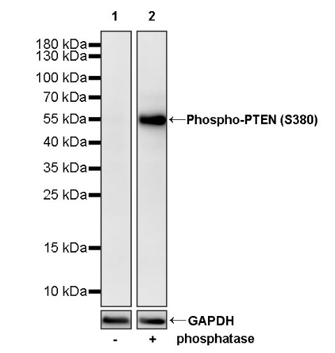
WB result of Phospho-PTEN (S380) Recombinant Rabbit mAb
Primary antibody: Phospho-PTEN (S380) Recombinant Rabbit mAb at 1/1000 dilution
Lane 1: HeLa whole cell lysate 20 µg
Lane 2: phosphatase treated HeLa whole cell 20 µg
Secondary antibody: Goat Anti-rabbit IgG, (H+L), HRP conjugated at 1/10000 dilution
Predicted MW: 47 kDa
Observed MW: 54 kDa
WB result of Phospho-PTEN (S380) Recombinant Rabbit mAb
Primary antibody: Phospho-PTEN (S380) Recombinant Rabbit mAb at 1/1000 dilution
Lane 1: NIH/3T3 whole cell lysate 20 µg
Lane 2: phosphatase treated HeLa whole cell 20 µg
Secondary antibody: Goat Anti-rabbit IgG, (H+L), HRP conjugated at 1/10000 dilution
Predicted MW: 47 kDa
Observed MW: 54 kDa
WB result of Phospho-PTEN (S380) Recombinant Rabbit mAb
Primary antibody: Phospho-PTEN (S380) Recombinant Rabbit mAb at 1/1000 dilution
Lane 1: C6 whole cell lysate 20 µg
Lane 2: phosphatase treated C6 whole cell 20 µg
Secondary antibody: Goat Anti-rabbit IgG, (H+L), HRP conjugated at 1/10000 dilution
Predicted MW: 47 kDa
Observed MW: 54 kDa
Flow cytometric analysis of 4% PFA fixed 90% methanol permeabilized HeLa (Human cervix adenocarcinoma epithelial cell) labelling Phospho-PTEN (S380) antibody at 1/500 dilution (0.1 μg) / (Red) compared with a Rabbit monoclonal IgG (Black) isotype control and an unlabelled control (cells without incubation with primary antibody and secondary antibody) (Blue). Goat Anti - Rabbit IgG Alexa Fluor® 488 was used as the secondary antibody.
Flow cytometric analysis of 4% PFA fixed 90% methanol permeabilized NIH/3T3 (Mouse embryonic fibroblast) labelling Phospho-PTEN (S380) antibody at 1/500 dilution (0.1 μg) / (Red) compared with a Rabbit monoclonal IgG (Black) isotype control and an unlabelled control (cells without incubation with primary antibody and secondary antibody) (Blue). Goat Anti - Rabbit IgG Alexa Fluor® 488 was used as the secondary antibody.
Phospho-PTEN (S380) Rabbit mAb at 1/50 dilution (1 µg) immunoprecipitating Phospho-PTEN (S380) in 0.4 mg HeLa whole cell lysate.
Western blot was performed on the immunoprecipitate using Phospho-PTEN (S380) Rabbit mAb at 1/1000 dilution.
Secondary antibody (HRP) for IP was used at 1/1000 dilution.
Lane 1: HeLa whole cell lysate 20 µg (Input)
Lane 2: Phospho-PTEN (S380) Rabbit mAb IP in HeLa whole cell lysate
Lane 3: Rabbit monoclonal IgG IP in HeLa whole cell lysate
Predicted MW: 47 kDa
Observed MW: 54 kDa
Dot blot result of Phospho-PTEN (S380) Recombinant Rabbit mAb
Lane 1: PTEN (S380) phospho peptide
Lane 2: PTEN unmodified peptide
Primary antibody: Phospho-PTEN (S380) Recombinant Rabbit mAb at 1/1000 dilution
Secondary antibody: Goat Anti-rabbit IgG, (H+L), HRP conjugated at 1/10000 dilution
ICC shows positive staining in HeLa cells. Anti- Phospho-PTEN (S380) antibody was used at 1/500 dilution (Green) and incubated overnight at 4°C. Goat polyclonal Antibody to Rabbit IgG - H&L (Alexa Fluor® 488) was used as secondary antibody at 1/1000 dilution. The cells were fixed with 4% PFA and permeabilized with 0.1% PBS-Triton X-100. Nuclei were counterstained with DAPI (Blue). Counterstain with tubulin (Red).
ICC shows positive staining in NIH/3T3 cells. Anti- Phospho-PTEN (S380) antibody was used at 1/500 dilution (Green) and incubated overnight at 4°C. Goat polyclonal Antibody to Rabbit IgG - H&L (Alexa Fluor® 488) was used as secondary antibody at 1/1000 dilution. The cells were fixed with 4% PFA and permeabilized with 0.1% PBS-Triton X-100. Nuclei were counterstained with DAPI (Blue). Counterstain with tubulin (Red).
 您现在的位置: