
PBS, 40% Glycerol, 0.05% BSA, 0.03% Proclin 300
12 months from date of receipt / reconstitution, -20 °C as supplied
| 应用 | 稀释度 |
|---|---|
| WB | 1:4000 |
| IP | 1:50 |
| IHC-P | 1:500 |
| ICC | 1:2000 |
| ICFCM | 1:200 |
CDK6 (Cyclin-dependent kinase 6) is a protein kinase that belongs to the CDK family, which plays a significant role in the cell cycle. It is involved in driving the cell cycle from the G1 phase into the S phase by forming complexes with D-type cyclins (D1, D2, and D3). CDK6, along with CDK4, phosphorylates the retinoblastoma (Rb) protein family, leading to the release of E2F transcription factors and enabling G1 to S phase progression and DNA synthesis. CDK6 is not only important for cell cycle regulation but also has kinase-independent functions, such as acting as a transcriptional regulator. This dual functionality makes CDK6 an attractive target for therapeutic intervention in various types of cancer, including hematopoietic malignancies, breast cancer, and melanoma.
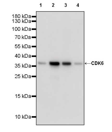
WB result of CDK6 Recombinant Rabbit mAb
Primary antibody: CDK6 Recombinant Rabbit mAb at 1/4000 dilution
Lane 1: HeLa whole cell lysate 20 µg
Lane 2: Jurkat whole cell lysate 20 µg
Lane 3: K562 whole cell lysate 20 µg
Lane 4: HEK-293 whole cell lysate 20 µg
Secondary antibody: Goat Anti-rabbit IgG, (H+L), HRP conjugated at 1/10000 dilution
Predicted MW: 37 kDa
Observed MW: 37 kDa
WB result of CDK6 Recombinant Rabbit mAb
Primary antibody: CDK6 Recombinant Rabbit mAb at 1/4000 dilution
Lane 1: mouse spleen lysate 20 µg
Secondary antibody: Goat Anti-rabbit IgG, (H+L), HRP conjugated at 1/10000 dilution
Predicted MW: 37 kDa
Observed MW: 37 kDa
WB result of CDK6 Recombinant Rabbit mAb
Primary antibody: CDK6 Recombinant Rabbit mAb at 1/4000 dilution
Lane 1: C6 whole cell lysate 20 µg
Secondary antibody: Goat Anti-rabbit IgG, (H+L), HRP conjugated at 1/10000 dilution
Predicted MW: 37 kDa
Observed MW: 37 kDa
Flow cytometric analysis of 4% PFA fixed 90% methanol permeabilized HeLa (Human cervix adenocarcinoma epithelial cell) labelling CDK6 zeta antibody at 1/200 dilution (1 μg)/ (Red) compared with a Rabbit monoclonal IgG (Black) isotype control and an unlabelled control (cells without incubation with primary antibody and secondary antibody) (Blue). Goat Anti - Rabbit IgG Alexa Fluor® 488 was used as the secondary antibody.
CDK6 Rabbit mAb at 1/200 dilution (1 µg) immunoprecipitating CDK6 in 0.4 mg K562 whole cell lysate.
Western blot was performed on the immunoprecipitate using CDK6 Rabbit mAb at 1/1000 dilution.
Secondary antibody (HRP) for IP was used at 1/1000 dilution.
Lane 1: K562 whole cell lysate 20 µg (Input)
Lane 2: CDK6 Rabbit mAb IP in K562 whole cell lysate
Lane 3: Rabbit monoclonal IgG IP in K562 whole cell lysate
Predicted MW: 37 kDa
Observed MW: 37 kDa
IHC shows positive staining in paraffin-embedded human tonsil. Anti- CDK6 antibody was used at 1/500 dilution, followed by a HRP Polymer for Mouse & Rabbit IgG (ready to use). Counterstained with hematoxylin. Heat mediated antigen retrieval with Tris/EDTA buffer pH9.0 was performed before commencing with IHC staining protocol.
IHC shows positive staining in paraffin-embedded human bladder cancer. Anti- CDK6 antibody was used at 1/500 dilution, followed by a HRP Polymer for Mouse & Rabbit IgG (ready to use). Counterstained with hematoxylin. Heat mediated antigen retrieval with Tris/EDTA buffer pH9.0 was performed before commencing with IHC staining protocol.
IHC shows positive staining in paraffin-embedded human endometrial cancer. Anti- CDK6 antibody was used at 1/500 dilution, followed by a HRP Polymer for Mouse & Rabbit IgG (ready to use). Counterstained with hematoxylin. Heat mediated antigen retrieval with Tris/EDTA buffer pH9.0 was performed before commencing with IHC staining protocol.
IHC shows positive staining in paraffin-embedded human lung cancer. Anti- CDK6 antibody was used at 1/500 dilution, followed by a HRP Polymer for Mouse & Rabbit IgG (ready to use). Counterstained with hematoxylin. Heat mediated antigen retrieval with Tris/EDTA buffer pH9.0 was performed before commencing with IHC staining protocol.
IHC shows positive staining in paraffin-embedded human ovarian cancer. Anti- CDK6 antibody was used at 1/500 dilution, followed by a HRP Polymer for Mouse & Rabbit IgG (ready to use). Counterstained with hematoxylin. Heat mediated antigen retrieval with Tris/EDTA buffer pH9.0 was performed before commencing with IHC staining protocol.
ICC shows positive staining in HeLa cells. Anti-CDK6 antibody was used at 1/2000 dilution (Green) and incubated overnight at 4°C. Goat polyclonal Antibody to Rabbit IgG - H&L (Alexa Fluor® 488) was used as secondary antibody at 1/1000 dilution. The cells were fixed with 100% ice-cold methanol and permeabilized with 0.1% PBS-Triton X-100. Nuclei were counterstained with DAPI (Blue). Counterstain with tubulin (Red).
 您现在的位置: