
PBS, 40% Glycerol, 0.05% BSA, 0.03% Proclin 300
12 months from date of receipt / reconstitution, -20 °C as supplied
| 应用 | 稀释度 |
|---|---|
| WB | 1:1000 |
| IP | 1:50 |
| IHC-P | 1:1000 |
CPT1A, or carnitine palmitoyltransferase 1A, is a crucial enzyme involved in the mitochondrial fatty acid oxidation (FAO) pathway, which is essential for energy homeostasis during fasting or prolonged exercise. It is responsible for the rate-limiting step of converting acyl-coenzyme as into acyl-carnitines, facilitating their transport into the mitochondria for oxidation. The regulation of CPT1A is complex and involves various genetic, epigenetic, physiological, and nutritional factors. In the context of cancer, CPT1A plays a significant role in tumor progression and metabolism. It has been found to be upregulated in various cancers, including lung cancer, and its increased expression is associated with poor prognosis. Targeting CPT1A has been proposed as a potential therapeutic strategy in cancer treatment. Inhibiting CPT1A can induce ferroptosis in cancer cells, including LCSCs, and enhance the efficacy of immune checkpoint therapy in lung cancer. Moreover, the combination of CPT1A inhibition with immunotherapy has shown synergistic effects in preclinical models, offering a new approach to overcome resistance to immunotherapy in lung cancer. Furthermore, CPT1A has been implicated in the metabolic reprogramming of cancer cells, promoting cell proliferation via nucleoside metabolism in nasopharyngeal carcinoma. It also plays a role in conferring cancer cell resistance to immune-mediated cytolytic killing.
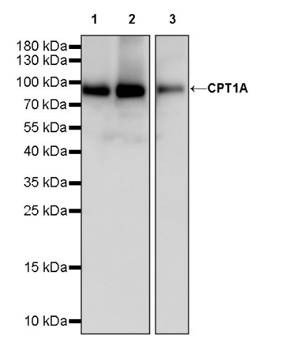
WB result of CPT1A Recombinant Rabbit mAb
Primary antibody: CPT1A Recombinant Rabbit mAb at 1/1000 dilution
Lane 1: HeLa whole cell lysate 20 µg
Lane 2: MCF7 whole cell lysate 20 µg
Lane 3: HepG2 whole cell lysate 20 µg
Secondary antibody: Goat Anti-rabbit IgG, (H+L), HRP conjugated at 1/10000 dilution
Predicted MW: 88 kDa
Observed MW: 90 kDa
CPT1A Rabbit mAb at 1/50 dilution (1 µg) immunoprecipitating CPT1A in 0.4 mg HeLa whole cell lysate.
Western blot was performed on the immunoprecipitate using CPT1A Rabbit mAb at 1/1000 dilution.
Secondary antibody (HRP) for IP was used at 1/1000 dilution.
Lane 1: HeLa whole cell lysate 20 µg (Input)
Lane 2: CPT1A Rabbit mAb IP in HeLa whole cell lysate
Lane 3: Rabbit monoclonal IgG IP in HeLa whole cell lysate
Predicted MW: 88 kDa
Observed MW: 90 kDa
IHC shows positive staining in paraffin-embedded human kidney. Anti- CPT1A antibody was used at 1/1000 dilution, followed by a HRP Polymer for Mouse & Rabbit IgG (ready to use). Counterstained with hematoxylin. Heat mediated antigen retrieval with Tris/EDTA buffer pH9.0 was performed before commencing with IHC staining protocol.
IHC shows positive staining in paraffin-embedded human liver. Anti- CPT1A antibody was used at 1/1000 dilution, followed by a HRP Polymer for Mouse & Rabbit IgG (ready to use). Counterstained with hematoxylin. Heat mediated antigen retrieval with Tris/EDTA buffer pH9.0 was performed before commencing with IHC staining protocol.
IHC shows positive staining in paraffin-embedded human endometrial cancer. Anti- CPT1A antibody was used at 1/1000 dilution, followed by a HRP Polymer for Mouse & Rabbit IgG (ready to use). Counterstained with hematoxylin. Heat mediated antigen retrieval with Tris/EDTA buffer pH9.0 was performed before commencing with IHC staining protocol.
IHC shows positive staining in paraffin-embedded human hepatocellular carcinoma. Anti- CPT1A antibody was used at 1/1000 dilution, followed by a HRP Polymer for Mouse & Rabbit IgG (ready to use). Counterstained with hematoxylin. Heat mediated antigen retrieval with Tris/EDTA buffer pH9.0 was performed before commencing with IHC staining protocol.
 您现在的位置: