
PBS, 40% Glycerol, 0.05% BSA, 0.03% Proclin 300
12 months from date of receipt / reconstitution, -20 °C as supplied
| 应用 | 稀释度 |
|---|---|
| WB | 1:1000 |
| IHC-P | 1:1000 |
| ICC | 1:500 |
eIF5A, or eukaryotic initiation factor 5A, is a protein involved in the process of translation within cells. It plays a role in the initiation, elongation, and termination phases of protein synthesis. eIF5A is unique in that it contains the rare amino acid hypusine, which is formed through a post-translational modification process involving the polyamine pathway. This factor is highly conserved across species and is implicated in various cellular functions beyond translation, such as cell proliferation, development, and response to stress. Recent research has shed light on the broader physiological roles of eIF5A, including its involvement in processes like ischemic tolerance, metabolic adaptation, aging, immune cell differentiation, and even in diseases where oxygen supply is disrupted, like organ transplantation, myocardial infarction, or stroke.
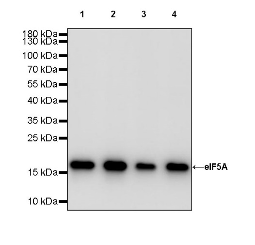
WB result of eIF5A Rabbit pAb
Primary antibody: eIF5A Rabbit pAb at 1/1000 dilution
Lane 1: Jurkat whole cell lysate 20 µg
Lane 2: HeLa whole cell lysate 20 µg
Lane 3: HepG2 whole cell lysate 20 µg
Lane 4: Caco-2 whole cell lysate 20 µg
Secondary antibody: Goat Anti-rabbit IgG, (H+L), HRP conjugated at 1/10000 dilution
Predicted MW: 17 kDa
Observed MW: 17 kDa
WB result of eIF5A Rabbit pAb
Primary antibody: eIF5A Rabbit pAb at 1/1000 dilution
Lane 1: NIH/3T3 whole cell lysate 20 µg
Lane 2: mouse heart lysate 20 µg
Secondary antibody: Goat Anti-rabbit IgG, (H+L), HRP conjugated at 1/10000 dilution
Predicted MW: 17 kDa
Observed MW: 17 kDa
WB result of eIF5A Rabbit pAb
Primary antibody: eIF5A Rabbit pAb at 1/1000 dilution
Lane 1: C6 whole cell lysate 20 µg
Lane 2: rat heart lysate 20 µg
Secondary antibody: Goat Anti-rabbit IgG, (H+L), HRP conjugated at 1/10000 dilution
Predicted MW: 17 kDa
Observed MW: 17 kDa
IHC shows positive staining in paraffin-embedded human tonsil. Anti- eIF5A antibody was used at 1/1000 dilution, followed by a HRP Polymer for Mouse & Rabbit IgG (ready to use). Counterstained with hematoxylin. Heat mediated antigen retrieval with Tris/EDTA buffer pH9.0 was performed before commencing with IHC staining protocol.
IHC shows positive staining in paraffin-embedded human stomach. Anti- eIF5A antibody was used at 1/1000 dilution, followed by a HRP Polymer for Mouse & Rabbit IgG (ready to use). Counterstained with hematoxylin. Heat mediated antigen retrieval with Tris/EDTA buffer pH9.0 was performed before commencing with IHC staining protocol.
IHC shows positive staining in paraffin-embedded human ovarian cancer. Anti- eIF5A antibody was used at 1/1000 dilution, followed by a HRP Polymer for Mouse & Rabbit IgG (ready to use). Counterstained with hematoxylin. Heat mediated antigen retrieval with Tris/EDTA buffer pH9.0 was performed before commencing with IHC staining protocol.
IHC shows positive staining in paraffin-embedded mouse kidney. Anti- eIF5A antibody was used at 1/1000 dilution, followed by a HRP Polymer for Mouse & Rabbit IgG (ready to use). Counterstained with hematoxylin. Heat mediated antigen retrieval with Tris/EDTA buffer pH9.0 was performed before commencing with IHC staining protocol.
IHC shows positive staining in paraffin-embedded rat stomach. Anti- eIF5A antibody was used at 1/1000 dilution, followed by a HRP Polymer for Mouse & Rabbit IgG (ready to use). Counterstained with hematoxylin. Heat mediated antigen retrieval with Tris/EDTA buffer pH9.0 was performed before commencing with IHC staining protocol.
ICC shows positive staining in HeLa cells. Anti- eIF5A antibody was used at 1/500 dilution (Green) and incubated overnight at 4°C. Goat polyclonal Antibody to Rabbit IgG - H&L (Alexa Fluor® 488) was used as secondary antibody at 1/1000 dilution. The cells were fixed with 4% PFA and permeabilized with 0.1% PBS-Triton X-100. Nuclei were counterstained with DAPI (Blue). Counterstain with tubulin (Red).
ICC shows positive staining in NIH/3T3 cells. Anti- eIF5A antibody was used at 1/500 dilution (Green) and incubated overnight at 4°C. Goat polyclonal Antibody to Rabbit IgG - H&L (Alexa Fluor® 488) was used as secondary antibody at 1/1000 dilution. The cells were fixed with 4% PFA and permeabilized with 0.1% PBS-Triton X-100. Nuclei were counterstained with DAPI (Blue). Counterstain with tubulin (Red).
 您现在的位置: