
PBS, 40% Glycerol, 0.05% BSA, 0.03% Proclin 300
12 months from date of receipt / reconstitution, -20 °C as supplied
| 应用 | 稀释度 |
|---|---|
| WB | 1:1000 |
| IP | 1:50 |
| IHC-P | 1:200 |
| ICC | 1:500 |
| IF | 1:100 |
p21Cip1 (alternatively p21Waf1), also known as cyclin-dependent kinase inhibitor 1 or CDK-interacting protein 1, is a cyclin-dependent kinase inhibitor (CKI) that is capable of inhibiting all cyclin/CDK complexes [PMID: 8259214], though is primarily associated with inhibition of CDK2 [PMID: 8259214, PMID: 8242751]. p21 represents a major target of p53 activity and thus is associated with linking DNA damage to cell cycle arrest [PMID: 9822382]. his protein is encoded by the CDKN1A gene located on chromosome 6 (6p21.2) in humans.
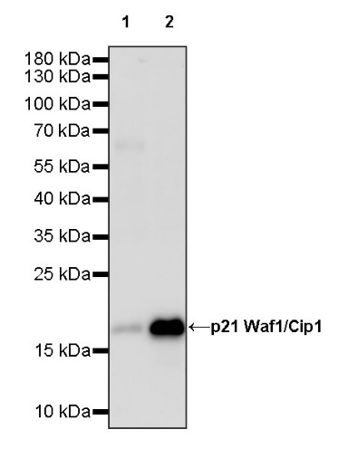
WB result of p21 Waf1/Cip1 Recombinant Rabbit mAb
Primary antibody: p21 Waf1/Cip1 Recombinant Rabbit mAb at 1/1000 dilution
Lane 1: HeLa whole cell lysate 20 µg
Lane 2: MCF7 whole cell lysate 20 µg
Weak expression: HeLa whole cell lysate
Secondary antibody: Goat Anti-rabbit IgG, (H+L), HRP conjugated at 1/10000 dilution
Predicted MW: 18 kDa
Observed MW: 18 kDa
p21 Waf1/Cip1 Rabbit mAb at 1/50 dilution (1 µg) immunoprecipitating p21 Waf1/Cip1 in 0.4 mg MCF7 whole cell lysate.
Western blot was performed on the immunoprecipitate using p21 Waf1/Cip1 Rabbit mAb at 1/1000 dilution.
Secondary antibody (HRP) for IP was used at 1/1000 dilution.
Lane 1: MCF7 whole cell lysate 20 µg (Input)
Lane 2: p21 Waf1/Cip1 Rabbit mAb IP in MCF7 whole cell lysate
Lane 3: Rabbit monoclonal IgG IP in MCF7 whole cell lysate
Predicted MW: 18 kDa
Observed MW: 18 kDa
IHC shows positive staining in paraffin-embedded human tonsil. Anti- p21 Waf1/Cip1 antibody was used at 1/200 dilution, followed by a HRP Polymer for Mouse & Rabbit IgG (ready to use). Counterstained with hematoxylin. Heat mediated antigen retrieval with Tris/EDTA buffer pH9.0 was performed before commencing with IHC staining protocol.
IHC shows positive staining in paraffin-embedded human transitional cell carcinoma. Anti- p21 Waf1/Cip1 antibody was used at 1/200 dilution, followed by a HRP Polymer for Mouse & Rabbit IgG (ready to use). Counterstained with hematoxylin. Heat mediated antigen retrieval with Tris/EDTA buffer pH9.0 was performed before commencing with IHC staining protocol.
IHC shows positive staining in paraffin-embedded human breast cancer. Anti- p21 Waf1/Cip1 antibody was used at 1/200 dilution, followed by a HRP Polymer for Mouse & Rabbit IgG (ready to use). Counterstained with hematoxylin. Heat mediated antigen retrieval with Tris/EDTA buffer pH9.0 was performed before commencing with IHC staining protocol.
IHC shows positive staining in paraffin-embedded human cervical squamous cell carcinoma. Anti- p21 Waf1/Cip1 antibody was used at 1/200 dilution, followed by a HRP Polymer for Mouse & Rabbit IgG (ready to use). Counterstained with hematoxylin. Heat mediated antigen retrieval with Tris/EDTA buffer pH9.0 was performed before commencing with IHC staining protocol.
IHC shows positive staining in paraffin-embedded human thyroid cancer. Anti- p21 Waf1/Cip1 antibody was used at 1/200 dilution, followed by a HRP Polymer for Mouse & Rabbit IgG (ready to use). Counterstained with hematoxylin. Heat mediated antigen retrieval with Tris/EDTA buffer pH9.0 was performed before commencing with IHC staining protocol.
ICC shows positive staining in MCF7 cells. Anti- p21 Waf1/Cip1 antibody was used at 1/500 dilution (Green) and incubated overnight at 4°C. Goat polyclonal Antibody to Rabbit IgG - H&L (Alexa Fluor® 488) was used as secondary antibody at 1/1000 dilution. The cells were fixed with 100% ice-cold methanol and permeabilized with 0.1% PBS-Triton X-100. Nuclei were counterstained with DAPI (Blue). Counterstain with tubulin (Red).
IF shows positive staining in paraffin-embedded human lung adenocarcinoma. Anti-p21 Waf1/Cip1 antibody was used at 1/100 dilution (Red) and incubated overnight at 4°C. Goat polyclonal Antibody to Rabbit IgG - H&L (Alexa Fluor® 594) was used as secondary antibody at 1/500 dilution. Counterstained with DAPI (Blue). Heat mediated antigen retrieval with EDTA buffer pH9.0 was performed before commencing with IF staining protocol.
IF shows positive staining in paraffin-embedded human bladder cancer. Anti-p21 Waf1/Cip1 antibody was used at 1/100 dilution (Red) and incubated overnight at 4°C. Goat polyclonal Antibody to Rabbit IgG - H&L (Alexa Fluor® 594) was used as secondary antibody at 1/500 dilution. Counterstained with DAPI (Blue). Heat mediated antigen retrieval with EDTA buffer pH9.0 was performed before commencing with IF staining protocol.
 您现在的位置: