
PBS, 40% Glycerol, 0.05% BSA, 0.03% Proclin 300
12 months from date of receipt / reconstitution, -20 °C as supplied
| 应用 | 稀释度 |
|---|---|
| ICC | 1:500 |
| IF | 1:200 |
| FCM | 1:2000 |
Mouse I-A/I-E refers to the murine major histocompatibility complex class II (MHC class II) molecules, which play a central role in the immune system by presenting antigens to T cells. The I-A and I-E molecules are expressed on the surface of antigen-presenting cells (APCs), such as B cells, macrophages, and dendritic cells, and are responsible for displaying peptides derived from extracellular proteins to CD4+ T cells. This interaction is crucial for initiating the adaptive immune response. Antibodies against mouse I-A/I-E can be used in various scientific applications, including flow cytometry, immunohistochemistry, and immunoprecipitation. These antibodies can target MHC class II molecules in different species, including mice, and are essential tools for research in immunology and cell biology.
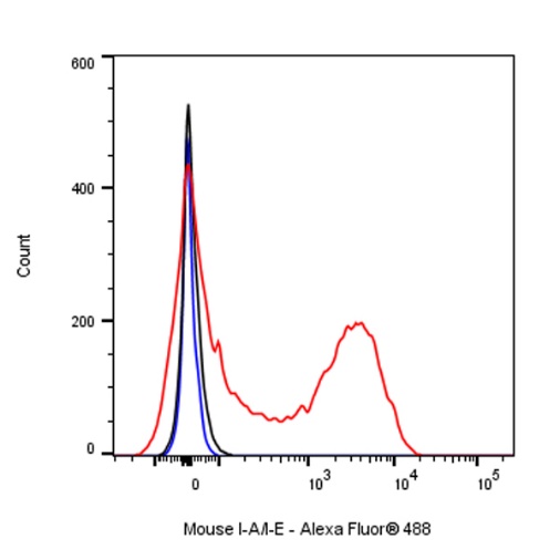
Flow cytometric analysis of C57BL/6 mouse splenocytes labelling Mouse I-A/I-E antibody at 1/2000 dilution (0.1 μg) / (Red) compared with a Rat monoclonal IgG (Black) isotype control and an unlabelled control (cells without incubation with primary antibody and secondary antibody) (Blue). Goat Anti - Rat IgG Alexa Fluor® 488 was used as the secondary antibody.
ICC shows positive staining in mouse splenocytes. Anti-Mouse I-A/I-E antibody was used at 1/500 dilution (Green) and incubated overnight at 4°C. Goat polyclonal Antibody to Rat IgG - H&L (Alexa Fluor® 488) was used as secondary antibody at 1/1000 dilution. The cells were fixed with 4% PFA and permeabilized with 0.1% PBS-Triton X-100. Nuclei were counterstained with DAPI (Blue). Counterstain with tubulin (Red).
IF shows positive staining in paraffin-embedded mouse spleen. Anti-Mouse I-A/I-E antibody was used at 1/200 dilution (Green) and incubated overnight at 4°C. Goat polyclonal Antibody to Rat IgG - H&L (Alexa Fluor® 488) was used as secondary antibody at 1/1000 dilution. Counterstained with DAPI (Blue). Heat mediated antigen retrieval with EDTA buffer pH9.0 was performed before commencing with IF staining protocol.
 您现在的位置: