
PBS, 40% Glycerol, 0.05% BSA, 0.03% Proclin 300
12 months from date of receipt / reconstitution, -20 °C as supplied
| 应用 | 稀释度 |
|---|---|
| WB | 1:1000 |
| IP | 1:50 |
SMAD3 is a protein intracellular signal transducer that is part of the TGF-β (transforming growth factor-beta) superfamily signaling pathway. When SMAD3 is phosphorylated at serine residues 423 and 425 (referred to as SMAD3 (phospho S423+S425)), it indicates activation of the TGF-β signaling cascade within a cell. This phosphorylation event typically occurs after SMAD3 binds to the activated TGF-β receptor, leading to its phosphorylation and subsequent interaction with SMAD4. Together, they accumulate in the nucleus where they regulate gene expression associated with various cellular processes, including cell growth, differentiation, and immune responses. In the context of disease research, understanding SMAD3 phosphorylation is particularly relevant in conditions such as cancer, where dysregulation of TGF-β signaling can contribute to tumor progression and metastasis. SMAD3 is also implicated in fibrotic diseases, cardiovascular diseases, and immune responses, making it a potential therapeutic target for these conditions.
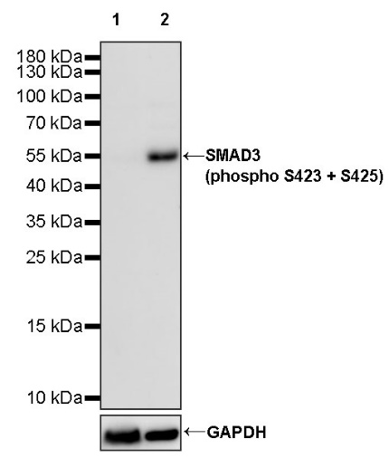
WB result of SMAD3 (phospho S423 + S425) Recombinant Rabbit mAb
Primary antibody: SMAD3 (phospho S423 + S425) Recombinant Rabbit mAb at 1/1000 dilution
Lane 1: untreated HT-1080 whole cell lysate 20 µg
Lane 2: HT-1080 treated with 10 ng/ml TGF-β for 30 minutes whole cell lysate 20 µg
Secondary antibody: Goat Anti-rabbit IgG, (H+L), HRP conjugated at 1/10000 dilution
Predicted MW: 48 kDa
Observed MW: 55 kDa
WB result of SMAD3 (phospho S423 + S425) Recombinant Rabbit mAb
Primary antibody: SMAD3 (phospho S423 + S425) Recombinant Rabbit mAb at 1/1000 dilution
Lane 1: untreated C2C12 whole cell lysate 20 µg
Lane 2: C2C12 treated with 10 ng/ml TGF-β for 30 minutes whole cell lysate 20 µg
Secondary antibody: Goat Anti-rabbit IgG, (H+L), HRP conjugated at 1/10000 dilution
Predicted MW: 48 kDa
Observed MW: 55 kDa
WB result of SMAD3 (phospho S423 + S425) Recombinant Rabbit mAb
Primary antibody: SMAD3 (phospho S423 + S425) Recombinant Rabbit mAb at 1/1000 dilution
Lane 1: untreated C6 whole cell lysate 20 µg
Lane 2: C6 treated with 10 ng/ml TGF-β for 30 minutes whole cell lysate 20 µg
Secondary antibody: Goat Anti-rabbit IgG, (H+L), HRP conjugated at 1/10000 dilution
Predicted MW: 48 kDa
Observed MW: 55 kDa
Smad3 (phospho S423 + S425) Recombinant Rabbit mAb at 1/50 dilution (1 µg) immunoprecipitating Smad3 (phospho S423 + S425) in 0.4 mg HT-1080+TGF-β(10 ng/ml, 30 min) whole cell lysate.
Western blot was performed on the immunoprecipitate using Smad3 (phospho S423 + S425) Recombinant Rabbit mAb at 1/1000 dilution.
Secondary antibody (HRP) for IP was used at 1/1000 dilution.
Lane 1: HT-1080+TGF-β(10 ng/ml, 30 min) whole cell lysate 20 µg (Input)
Lane 2: Smad3 (phospho S423 + S425) Recombinant Rabbit mAb IP in HT-1080+TGF-β(10 ng/ml, 30 min) whole cell lysate
Lane 3: Rabbit monoclonal IgG IP in HT-1080+TGF-β(10 ng/ml, 30 min) whole cell lysate
Predicted MW: 48 kDa
Observed MW: 55 kDa
Exposure time: 20 s
 您现在的位置: