
12 months from date of receipt / reconstitution, -20 °C as supplied
| 应用 | 稀释度 |
|---|---|
| WB | 1:1000 |
| IP | 1:50 |
| IHC-P | 1:1000 |
| ICC | 1:500 |
| ICFCM | 1:500 |
KAP-1, also known as KRAB-associated protein 1 or TIF1β (transcriptional intermediary factor 1β), is a multifunctional protein involved in a variety of cellular processes, including transcriptional regulation, DNA repair, and maintenance of genomic integrity. KAP-1 functions as a transcriptional corepressor by interacting with KRAB-ZNFs, which recruit it to specific genomic loci. Once bound, KAP-1 can influence the epigenetic state of the targeted DNA, leading to transcriptional repression through the recruitment of chromatin-modifying enzymes such as histone deacetylases (HDACs) and histone methyltransferases (HMTs) like SETDB1. This results in the formation of repressive chromatin marks, such as H3K9me3, which contribute to the establishment of a heterochromatic environment. In addition to its role in transcriptional regulation, KAP-1 has been implicated in the maintenance of pluripotency in stem cells and is required for terminal differentiation of mouse embryonic stem cells. It is also associated with promoting and inhibiting differentiation of various adult cell types, suggesting a complex role in cellular development and differentiation.
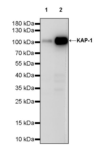
WB result of KAP-1 Recombinant Rabbit mAb
Primary antibody: KAP-1 Recombinant Rabbit mAb at 1/1000 dilution
Lane 1: A431 whole cell lysate 20 µg
Lane 2: MCF7 whole cell lysate 20 µg
Secondary antibody: Goat Anti-rabbit IgG, (H+L), HRP conjugated at 1/10000 dilution
Predicted MW: 89 kDa
Observed MW: 105 kDa
Flow cytometric analysis of 4% PFA fixed 90% methanol permeabilized HeLa (Human cervix adenocarcinoma epithelial cell) labelling KAP-1 antibody at 1/500 dilution (0.1 μg)/ (Red) compared with a Rabbit monoclonal IgG (Black) isotype control and an unlabelled control (cells without incubation with primary antibody and secondary antibody) (Blue). Goat Anti - Rabbit IgG Alexa Fluor® 488 was used as the secondary antibody.
KAP-1 Rabbit mAb at 1/50 dilution (1 µg) immunoprecipitating KAP-1 in 0.4 mg HeLa whole cell lysate.
Western blot was performed on the immunoprecipitate using KAP-1 Rabbit mAb at 1/1000 dilution.
Secondary antibody (HRP) for IP was used at 1/1000 dilution.
Lane 1: HeLa whole cell lysate 20 µg (Input)
Lane 2: KAP-1 Rabbit mAb IP in HeLa whole cell lysate
Lane 3: Rabbit monoclonal IgG IP in HeLa whole cell lysate
Predicted MW: 89 kDa
Observed MW: 105 kDa
IHC shows positive staining in paraffin-embedded human colon. Anti- KAP-1 antibody was used at 1/1000 dilution, followed by a HRP Polymer for Mouse & Rabbit IgG (ready to use). Counterstained with hematoxylin. Heat mediated antigen retrieval with Tris/EDTA buffer pH9.0 was performed before commencing with IHC staining protocol.
IHC shows positive staining in paraffin-embedded human kidney. Anti- KAP-1 antibody was used at 1/1000 dilution, followed by a HRP Polymer for Mouse & Rabbit IgG (ready to use). Counterstained with hematoxylin. Heat mediated antigen retrieval with Tris/EDTA buffer pH9.0 was performed before commencing with IHC staining protocol.
IHC shows positive staining in paraffin-embedded human breast cancer. Anti- KAP-1 antibody was used at 1/1000 dilution, followed by a HRP Polymer for Mouse & Rabbit IgG (ready to use). Counterstained with hematoxylin. Heat mediated antigen retrieval with Tris/EDTA buffer pH9.0 was performed before commencing with IHC staining protocol.
IHC shows positive staining in paraffin-embedded human colon cancer. Anti- KAP-1 antibody was used at 1/1000 dilution, followed by a HRP Polymer for Mouse & Rabbit IgG (ready to use). Counterstained with hematoxylin. Heat mediated antigen retrieval with Tris/EDTA buffer pH9.0 was performed before commencing with IHC staining protocol.
ICC shows positive staining in HeLa cells. Anti-KAP-1 antibody was used at 1/500 dilution (Green) and incubated overnight at 4°C. Goat polyclonal Antibody to Rabbit IgG - H&L (Alexa Fluor® 488) was used as secondary antibody at 1/1000 dilution. The cells were fixed with 4% PFA and permeabilized with 0.1% PBS-Triton X-100. Nuclei were counterstained with DAPI (Blue). Counterstain with tubulin (Red).
 您现在的位置: