
PBS, 40% Glycerol, 0.05% BSA, 0.03% Proclin 300
12 months from date of receipt / reconstitution, -20 °C as supplied
| 应用 | 稀释度 |
|---|---|
| WB | 1:4000 |
| IHC-P | 1:250 |
| ICC | 1:2000 |
| ICFCM | 1:2000 |
The BRAF protein is a serine/threonine-specific protein kinase that belongs to the RAF family of kinases, which are crucial components of the mitogen-activated protein kinase (MAPK) signaling pathway. This pathway, also known as the ERK signaling cascade, is involved in regulating a range of cellular activities including cell growth, differentiation, and survival. The BRAF protein plays a pivotal role in transducing signals from cell surface receptors to the nucleus, thereby influencing cellular behavior. Under normal conditions, the activation of BRAF is tightly regulated. However, when mutations occur in the BRAF gene, the resulting BRAF protein can become constitutively active, leading to uncontrolled signaling and contributing to the development and progression of cancer. The most well-known mutation is the BRAF V600E mutation, where a valine at position 600 is substituted by a glutamic acid. This mutation causes the BRAF protein to be continuously active, leading to increased cell proliferation and resistance to cell death.
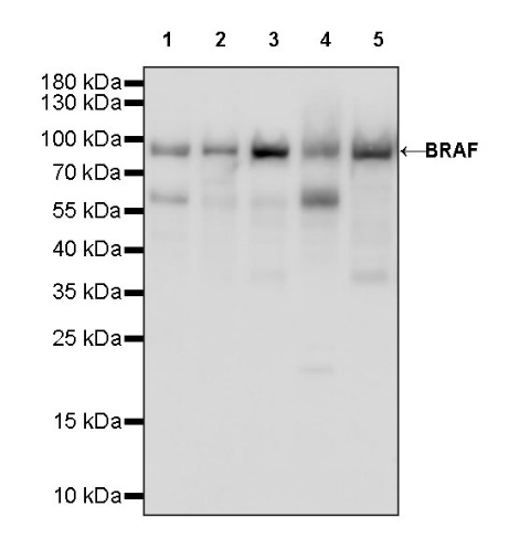
WB result of BRAF Recombinant Rabbit mAb
Primary antibody: BRAF Recombinant Rabbit mAb at 1/4000 dilution
Lane 1: HCT 116 whole cell lysate 20 µg
Lane 2: Caco-2 whole cell lysate 20 µg
Lane 3: 293T whole cell lysate 20 µg
Lane 4: SK-MEL-28 whole cell lysate 20 µg
Lane 5: K562 whole cell lysate 20 µg
Secondary antibody: Goat Anti-rabbit IgG, (H+L), HRP conjugated at 1/10000 dilution
Predicted MW: 84 kDa
Observed MW: 90 kDa
WB result of BRAF Recombinant Rabbit mAb
Primary antibody: BRAF Recombinant Rabbit mAb at 1/4000 dilution
Lane 1: A20 whole cell lysate 20 µg
Lane 2: mouse brain lysate 20 µg
Secondary antibody: Goat Anti-rabbit IgG, (H+L), HRP conjugated at 1/10000 dilution
Predicted MW: 84 kDa
Observed MW: 90 kDa
WB result of BRAF Recombinant Rabbit mAb
Primary antibody: BRAF Recombinant Rabbit mAb at 1/4000 dilution
Lane 1: PC-12 whole cell lysate 20 µg
Secondary antibody: Goat Anti-rabbit IgG, (H+L), HRP conjugated at 1/10000 dilution
Predicted MW: 84 kDa
Observed MW: 90 kDa
Flow cytometric analysis of 4% PFA fixed 90% methanol permeabilized HCT 116 (Human colorectal carcinoma epithelial cell) labelling BRAF antibody at 1/2000 dilution (0.1 μg) / (Red) compared with a Rabbit monoclonal IgG (Black) isotype control and an unlabelled control (cells without incubation with primary antibody and secondary antibody) (Blue). Goat Anti - Rabbit IgG Alexa Fluor® 488 was used as the secondary antibody.
IHC shows positive staining in paraffin-embedded human breast cancer. Anti-BRAF antibody was used at 1/250 dilution, followed by a HRP Polymer for Mouse & Rabbit IgG (ready to use). Counterstained with hematoxylin. Heat mediated antigen retrieval with Tris/EDTA buffer pH9.0 was performed before commencing with IHC staining protocol.
IHC shows positive staining in paraffin-embedded human gastric cancer. Anti-BRAF antibody was used at 1/250 dilution, followed by a HRP Polymer for Mouse & Rabbit IgG (ready to use). Counterstained with hematoxylin. Heat mediated antigen retrieval with Tris/EDTA buffer pH9.0 was performed before commencing with IHC staining protocol.
IHC shows positive staining in paraffin-embedded human ovarian cancer. Anti-BRAF antibody was used at 1/250 dilution, followed by a HRP Polymer for Mouse & Rabbit IgG (ready to use). Counterstained with hematoxylin. Heat mediated antigen retrieval with Tris/EDTA buffer pH9.0 was performed before commencing with IHC staining protocol.
IHC shows positive staining in paraffin-embedded mouse liver. Anti-BRAF antibody was used at 1/250 dilution, followed by a HRP Polymer for Mouse & Rabbit IgG (ready to use). Counterstained with hematoxylin. Heat mediated antigen retrieval with Tris/EDTA buffer pH9.0 was performed before commencing with IHC staining protocol.
IHC shows positive staining in paraffin-embedded rat kidney. Anti-BRAF antibody was used at 1/250 dilution, followed by a HRP Polymer for Mouse & Rabbit IgG (ready to use). Counterstained with hematoxylin. Heat mediated antigen retrieval with Tris/EDTA buffer pH9.0 was performed before commencing with IHC staining protocol.
IHC shows positive staining in paraffin-embedded human bladder cancer. Anti-BRAF antibody was used at 1/250 dilution, followed by a HRP Polymer for Mouse & Rabbit IgG (ready to use). Counterstained with hematoxylin. Heat mediated antigen retrieval with Tris/EDTA buffer pH9.0 was performed before commencing with IHC staining protocol.
ICC shows positive staining in HCT 116 cells. Anti-BRAF antibody was used at 1/2000 dilution (Green) and incubated overnight at 4°C. Goat polyclonal Antibody to Rabbit IgG - H&L (Alexa Fluor® 488) was used as secondary antibody at 1/1000 dilution. The cells were fixed with 4% PFA and permeabilized with 0.1% PBS-Triton X-100. Nuclei were counterstained with DAPI (Blue). Counterstain with tubulin (Red).
 您现在的位置: