
PBS, 40% Glycerol, 0.05% BSA, 0.03% Proclin 300
12 months from date of receipt / reconstitution, -20 °C as supplied
| 应用 | 稀释度 |
|---|---|
| WB | 1:1000 |
| IP | 1:50 |
| IHC-P | 1:500 |
| ICC | 1:500 |
Nucleolin, also known as C23 or NCL, is a multifunctional phosphoprotein that is predominantly found in the nucleolus of eukaryotic cells. It plays a crucial role in the biogenesis of ribosomes, including the processing of rRNA and the assembly of ribosomal subunits. Nucleolin is also involved in DNA replication, cell proliferation, and transcriptional regulation. In addition to its nuclear functions, nucleolin has been found on the cell surface, where it serves as a receptor for various ligands, such as growth factors and viruses. This cell surface expression of nucleolin is particularly significant in cancer cells and has been associated with poor prognosis. Nucleolin's presence on the cell surface also plays a critical role in modulating the cell cycle, cell proliferation, and apoptosis. Nucleolin has been identified as a cellular receptor for human respiratory syncytial virus, and it is also a target for nucleic acid-based adjuvants, which are important for vaccine development. Moreover, nucleolin's interaction with G-quadruplex structures within nucleic acids is a potential target for therapeutic interventions in diseases such as cancer and viral infections.
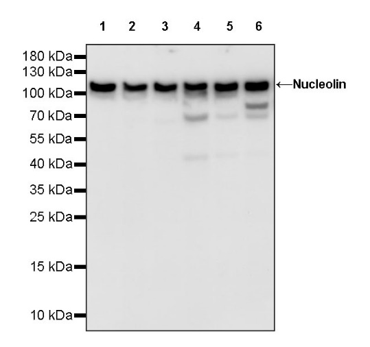
WB result of Nucleolin Rabbit pAb
Primary antibody: Nucleolin Rabbit pAb at 1/1000 dilution
Lane 1: HeLa whole cell lysate 20 µg
Lane 2: MCF7 whole cell lysate 20 µg
Lane 3: K562 whole cell lysate 20 µg
Lane 4: Jurkat whole cell lysate 20 µg
Lane 5: A431 whole cell lysate 20 µg
Lane 6: HT-29 whole cell lysate 20 µg
Secondary antibody: Goat Anti-rabbit IgG, (H+L), HRP conjugated at 1/10000 dilution
Predicted MW: 76 kDa
Observed MW: 110 kDa
WB result of Nucleolin Rabbit pAb
Primary antibody: Nucleolin Rabbit pAb at 1/1000 dilution
Lane 1: NIH/3T3 whole cell lysate 20 µg
Lane 2: Neuro-2a whole cell lysate 20 µg
Secondary antibody: Goat Anti-rabbit IgG, (H+L), HRP conjugated at 1/10000 dilution
Predicted MW: 76 kDa
Observed MW: 110 kDa
WB result of Nucleolin Rabbit pAb
Primary antibody: Nucleolin Rabbit pAb at 1/1000 dilution
Lane 1: PC-12 whole cell lysate 20 µg
Lane 2: C6 whole cell lysate 20 µg
Secondary antibody: Goat Anti-rabbit IgG, (H+L), HRP conjugated at 1/10000 dilution
Predicted MW: 76 kDa
Observed MW: 110 kDa
Flow cytometric analysis of 4% PFA fixed 90% methanol permeabilized HeLa (Human cervix adenocarcinoma epithelial cell) labelling Nucleolin antibody at 1/500 dilution (0.1 μg) / (Red) compared with a Rabbit monoclonal IgG (Black) isotype control and an unlabelled control (cells without incubation with primary antibody and secondary antibody) (Blue). Goat Anti - Rabbit IgG Alexa Fluor® 488 was used as the secondary antibody.
Nucleolin Rabbit pAb at 1/50 dilution (1 µg) immunoprecipitating Nucleolin in 0.4 mg HeLa whole cell lysate.
Western blot was performed on the immunoprecipitate using Nucleolin Rabbit pAb at 1/1000 dilution.
Secondary antibody (HRP) for IP was used at 1/1000 dilution.
Lane 1: HeLa whole cell lysate 20 µg (Input)
Lane 2: Nucleolin Rabbit pAb IP in HeLa whole cell lysate
Lane 3: Rabbit monoclonal IgG IP in HeLa whole cell lysate
Predicted MW: 76 kDa
Observed MW: 110 kDa
IHC shows positive staining in paraffin-embedded human colon. Anti- Nucleolin antibody was used at 1/500 dilution, followed by a HRP Polymer for Mouse & Rabbit IgG (ready to use). Counterstained with hematoxylin. Heat mediated antigen retrieval with Tris/EDTA buffer pH9.0 was performed before commencing with IHC staining protocol.
IHC shows positive staining in paraffin-embedded human kidney. Anti- Nucleolin antibody was used at 1/500 dilution, followed by a HRP Polymer for Mouse & Rabbit IgG (ready to use). Counterstained with hematoxylin. Heat mediated antigen retrieval with Tris/EDTA buffer pH9.0 was performed before commencing with IHC staining protocol.
IHC shows positive staining in paraffin-embedded human breast cancer. Anti- Nucleolin antibody was used at 1/500 dilution, followed by a HRP Polymer for Mouse & Rabbit IgG (ready to use). Counterstained with hematoxylin. Heat mediated antigen retrieval with Tris/EDTA buffer pH9.0 was performed before commencing with IHC staining protocol.
IHC shows positive staining in paraffin-embedded human lung adenocarcinoma. Anti- Nucleolin antibody was used at 1/500 dilution, followed by a HRP Polymer for Mouse & Rabbit IgG (ready to use). Counterstained with hematoxylin. Heat mediated antigen retrieval with Tris/EDTA buffer pH9.0 was performed before commencing with IHC staining protocol.
IHC shows positive staining in paraffin-embedded human ovarian cancer. Anti- Nucleolin antibody was used at 1/500 dilution, followed by a HRP Polymer for Mouse & Rabbit IgG (ready to use). Counterstained with hematoxylin. Heat mediated antigen retrieval with Tris/EDTA buffer pH9.0 was performed before commencing with IHC staining protocol.
IHC shows positive staining in paraffin-embedded human stomach. Anti- Nucleolin antibody was used at 1/500 dilution, followed by a HRP Polymer for Mouse & Rabbit IgG (ready to use). Counterstained with hematoxylin. Heat mediated antigen retrieval with Tris/EDTA buffer pH9.0 was performed before commencing with IHC staining protocol.
IHC shows positive staining in paraffin-embedded mouse colon. Anti- Nucleolin antibody was used at 1/500 dilution, followed by a HRP Polymer for Mouse & Rabbit IgG (ready to use). Counterstained with hematoxylin. Heat mediated antigen retrieval with Tris/EDTA buffer pH9.0 was performed before commencing with IHC staining protocol.
IHC shows positive staining in paraffin-embedded rat stomach. Anti- Nucleolin antibody was used at 1/500 dilution, followed by a HRP Polymer for Mouse & Rabbit IgG (ready to use). Counterstained with hematoxylin. Heat mediated antigen retrieval with Tris/EDTA buffer pH9.0 was performed before commencing with IHC staining protocol.
ICC shows positive staining in HeLa cells. Anti- Nucleolin antibody was used at 1/500 dilution (Green) and incubated overnight at 4°C. Goat polyclonal Antibody to Rabbit IgG - H&L (Alexa Fluor® 488) was used as secondary antibody at 1/1000 dilution. The cells were fixed with 4% PFA and permeabilized with 0.1% PBS-Triton X-100. Nuclei were counterstained with DAPI (Blue). Counterstain with tubulin (Red).
 您现在的位置: