
12 months from date of receipt / reconstitution, -20 °C as supplied
| 应用 | 稀释度 |
|---|---|
| WB | 1:1000 |
| ICC | 1:500 |
| ICFCM | 1:500 |
The CDK4 protein, or cyclin-dependent kinase 4, is a crucial regulator of the cell cycle, particularly in the G1 phase. It is a protein kinase that, in complex with cyclin D, phosphorylates the retinoblastoma tumor suppressor protein (pRb), leading to cell cycle progression from the G1 phase to the S phase. This process is essential for cell growth and division. CDK4 is often dysregulated in cancer, with overexpression or hyperactivity contributing to uncontrolled cell proliferation. Mutations in CDK4 can lead to its constitutive activation, which has been implicated in various types of cancer, including melanoma, glioblastoma, and breast cancer. The CDK4/6 inhibitors, such as palbociclib, ribociclib, and abemaciclib, have been developed as targeted therapies to treat certain breast cancers by inhibiting the activity of CDK4/6, thereby blocking cell cycle progression.
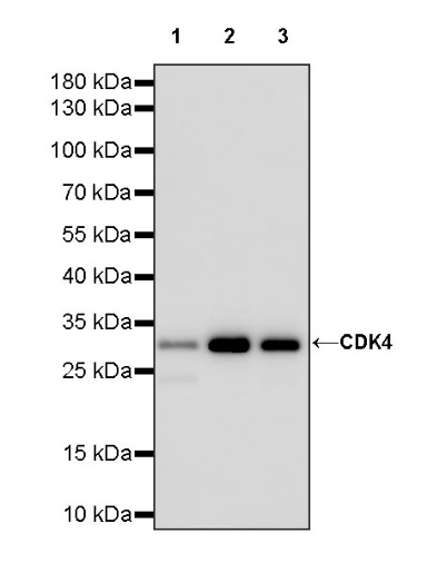
WB result of CDK4 Recombinant Rabbit mAb
Primary antibody: CDK4 Recombinant Rabbit mAb at 1/1000 dilution
Lane 1: Jurkat whole cell lysate 20 µg
Lane 2: HeLa whole cell lysate 20 µg
Lane 3: MCF7 whole cell lysate 20 µg
Secondary antibody: Goat Anti-rabbit IgG, (H+L), HRP conjugated at 1/10000 dilution
Predicted MW: 34 kDa
Observed MW: 30 kDa
Flow cytometric analysis of 4% PFA fixed 90% methanol permeabilized HeLa (Human cervix adenocarcinoma epithelial cell) labelling CDK4 antibody at 1/500 dilution (0.1 μg) / (Red) compared with a Rabbit monoclonal IgG (Black) isotype control and an unlabelled control (cells without incubation with primary antibody and secondary antibody) (Blue). Goat Anti - Rabbit IgG Alexa Fluor® 488 was used as the secondary antibody.
Flow cytometric analysis of 4% PFA fixed 90% methanol permeabilized MCF7 (Human breast adenocarcinoma epithelial cell) labelling CDK4 antibody at 1/500 dilution (0.1 μg) / (Red) compared with a Rabbit monoclonal IgG (Black) isotype control and an unlabelled control (cells without incubation with primary antibody and secondary antibody) (Blue). Goat Anti - Rabbit IgG Alexa Fluor® 488 was used as the secondary antibody.
ICC shows positive staining in HeLa cells. Anti-CDK4 antibody was used at 1/500 dilution (Green) and incubated overnight at 4°C. Goat polyclonal Antibody to Rabbit IgG - H&L (Alexa Fluor® 488) was used as secondary antibody at 1/1000 dilution. The cells were fixed with 100% ice-cold methanol and permeabilized with 0.1% PBS-Triton X-100. Nuclei were counterstained with DAPI (Blue). Counterstain with tubulin (Red).
ICC shows positive staining in MCF7 cells. Anti-CDK4 antibody was used at 1/500 dilution (Green) and incubated overnight at 4°C. Goat polyclonal Antibody to Rabbit IgG - H&L (Alexa Fluor® 488) was used as secondary antibody at 1/1000 dilution. The cells were fixed with 100% ice-cold methanol and permeabilized with 0.1% PBS-Triton X-100. Nuclei were counterstained with DAPI (Blue). Counterstain with tubulin (Red).
 您现在的位置: