
PBS, 40% Glycerol, 0.05% BSA, 0.03% Proclin 300
12 months from date of receipt / reconstitution, -20 °C as supplied
| 应用 | 稀释度 |
|---|---|
| Dot Blot | 1:1000 |
| WB | 1:2000 |
| IP | 1:50 |
| IHC-P | 1:500-1:2000 |
| ChIP | 1:20~1:50 |
Histone H4 acetylation at lysine 5 (H4K5ac) is a post-translational modification that plays a significant role in chromatin structure and function. This modification is catalyzed by histone acetyltransferases (HATs), which transfer an acetyl group from acetyl-coenzyme A (acetyl-CoA) to the ε-amino group of the lysine residue on histone H4. The acetylation of histone H4 at lysine 5 is crucial for various cellular processes, including gene activation, DNA replication, and cell cycle progression. It is involved in providing a more open chromatin structure, which is generally permissive for the binding of transcription factors and the assembly of the transcription machinery. Additionally, H4K5ac has been linked to the deposition of the histone H3 variant CENP-A into centromeres, which is essential for proper chromosome segregation during cell division.
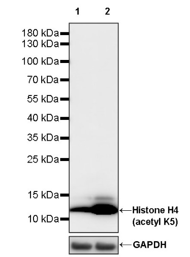
WB result of Histone H4 (acetyl K5) Recombinant Rabbit mAb
Primary antibody: Histone H4 (acetyl K5) Recombinant Rabbit mAb at 1/2000 dilution
Lane 1: untreated HeLa whole cell lysate 20 µg
Lane 2: HeLa treated with 500 ng/ml TSA for 4 hours whole cell lysate 20 µg
Secondary antibody: Goat Anti-rabbit IgG, (H+L), HRP conjugated at 1/10000 dilution
Predicted MW: 11 kDa
Observed MW: 11 kDa
WB result of Histone H4 (acetyl K5) Recombinant Rabbit mAb
Primary antibody: Histone H4 (acetyl K5) Recombinant Rabbit mAb at 1/2000 dilution
Lane 1: untreated NIH/3T3 whole cell lysate 20 µg
Lane 2: NIH/3T3 treated with 500 ng/ml TSA for 4 hours whole cell lysate 20 µg
Secondary antibody: Goat Anti-rabbit IgG, (H+L), HRP conjugated at 1/10000 dilution
Predicted MW: 11 kDa
Observed MW: 11 kDa
Histone H4 (acetyl K5) Rabbit pAb at 1/50 dilution (1 µg) immunoprecipitating Histone H4 (acetyl K5) in 0.4 mg NIH/3T3 treated with 500 ng/ml TSA for 4 hours whole cell lysate.
Western blot was performed on the immunoprecipitate using Histone H4 (acetyl K5) Rabbit pAb at 1/1000 dilution.
Secondary antibody (HRP) for IP was used at 1/1000 dilution.
Lane 1: NIH/3T3 treated with 500 ng/ml TSA for 4 hours whole cell lysate 5 µg (Input)
Lane 2: Histone H4 (acetyl K5) Rabbit pAb IP in NIH/3T3 treated with 500 ng/ml TSA for 4 hours whole cell lysate
Lane 3: Rabbit monoclonal IgG IP in NIH/3T3 treated with 500 ng/ml TSA for 4 hours whole cell lysate
Predicted MW: 11 kDa
Observed MW: 11 kDa
Dot blot result of Histone H4 (acetyl K5) Recombinant Rabbit mAb
Lane 1: H4K5ac modified peptide
Lane 2: H4K5 unmodified peptide
Lane 3: H2AK5ac modified peptide
Primary antibody: Histone H4 (acetyl K5) Recombinant Rabbit mAb at 1/1000 dilution
Secondary antibody: Goat Anti-rabbit IgG, (H+L), HRP conjugated at 1/10000 dilution
IHC shows positive staining in paraffin-embedded human colon. Anti- Histone H4 (acetyl K5) antibody was used at 1/500 dilution, followed by a HRP Polymer for Mouse & Rabbit IgG (ready to use). Counterstained with hematoxylin. Heat mediated antigen retrieval with Tris/EDTA buffer pH9.0 was performed before commencing with IHC staining protocol.
IHC shows positive staining in paraffin-embedded human kidney. Anti- Histone H4 (acetyl K5) antibody was used at 1/500 dilution, followed by a HRP Polymer for Mouse & Rabbit IgG (ready to use). Counterstained with hematoxylin. Heat mediated antigen retrieval with Tris/EDTA buffer pH9.0 was performed before commencing with IHC staining protocol.
IHC shows positive staining in paraffin-embedded human testis. Anti- Histone H4 (acetyl K5) antibody was used at 1/500 dilution, followed by a HRP Polymer for Mouse & Rabbit IgG (ready to use). Counterstained with hematoxylin. Heat mediated antigen retrieval with Tris/EDTA buffer pH9.0 was performed before commencing with IHC staining protocol.
IHC shows positive staining in paraffin-embedded mouse lung. Anti- Histone H4 (acetyl K5) antibody was used at 1/2000 dilution, followed by a HRP Polymer for Mouse & Rabbit IgG (ready to use). Counterstained with hematoxylin. Heat mediated antigen retrieval with Tris/EDTA buffer pH9.0 was performed before commencing with IHC staining protocol.
IHC shows positive staining in paraffin-embedded mouse testis. Anti- Histone H4 (acetyl K5) antibody was used at 1/2000 dilution, followed by a HRP Polymer for Mouse & Rabbit IgG (ready to use). Counterstained with hematoxylin. Heat mediated antigen retrieval with Tris/EDTA buffer pH9.0 was performed before commencing with IHC staining protocol.
IHC shows positive staining in paraffin-embedded rat colon. Anti- Histone H4 (acetyl K5) antibody was used at 1/2000 dilution, followed by a HRP Polymer for Mouse & Rabbit IgG (ready to use). Counterstained with hematoxylin. Heat mediated antigen retrieval with Tris/EDTA buffer pH9.0 was performed before commencing with IHC staining protocol.
IHC shows positive staining in paraffin-embedded rat testis. Anti- Histone H4 (acetyl K5) antibody was used at 1/2000 dilution, followed by a HRP Polymer for Mouse & Rabbit IgG (ready to use). Counterstained with hematoxylin. Heat mediated antigen retrieval with Tris/EDTA buffer pH9.0 was performed before commencing with IHC staining protocol.
Chromatin immunoprecipitation (ChIP) was
performed on HeLa+TSA (500ng/ml,4h) (+) cells
cross - linked with 1% formaldehyde for 10 min,
then chromatin was fragmented by sonication.
Parallel reactions used Histone H4 (acetyl K5)
Recombinant Rabbit mAb (S-1464-112) and Rabbit
mAb IgG Isotype Control (SDT-R173) at 1:50 for
immunoprecipitation.
Post - immunoprecipitation, both samples
were washed, eluted, and cross - links
reversed. Purified DNA was analyzed by qPCR.
qPCR (%input: immunoprecipitated DNA/input DNA)
showed the enrichment of RPL30, GAPDH, MYOD1, AFM,
SAT-α and SAT-2 in Histone H4 (acetyl K5) Recombinant
Rabbit mAb (S-1464-112)-immunoprecipitated sample.
 您现在的位置: