
12 months from date of receipt / reconstitution, -20 °C as supplied
| 应用 | 稀释度 |
|---|---|
| WB | 1:1000 |
| IP | 1:50 |
| IHC-P | 1:500 |
Growth differentiation factor-15 (GDF-15), also known as macrophage inhibitory cytokine-1 (MIC-1), is a member of the transforming growth factor-β (TGF-β) superfamily. It is a stress-responsive cytokine that is produced by various cell types including macrophages, vascular smooth muscle cells, cardiomyocytes, adipocytes, and endothelial cells. GDF-15 is typically expressed at low levels in most tissues but can be significantly induced under conditions of tissue injury, hypoxia, and inflammatory cytokine responses. It plays a role in various biological processes such as organ growth, differentiation, development, and cellular repair. Notably, GDF-15 has been identified as a biomarker for disease prognosis, particularly in cardiovascular diseases, cancer, and metabolic disorders. The cytokine has also been implicated in immune modulation and has been proposed as a potential target for therapeutic intervention in conditions like obesity, diabetes, and cardiovascular diseases. Its role as an immune checkpoint and its potential as a target for cancer immunotherapy are areas of active research.
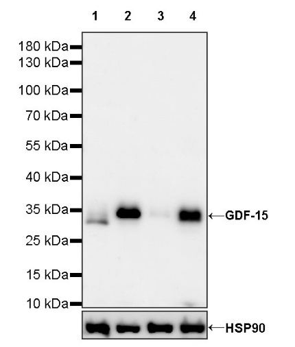
WB result of GDF-15 Recombinant Rabbit mAb
Primary antibody: GDF-15 Recombinant Rabbit mAb at 1/1000 dilution
Lane 1: untreated HT-1080 whole cell lysate 20 µg
Lane 2: HT-1080 treated with 200 ng/ml TPA for 24 hours whole cell lysate 20 µg
Lane 3: HepG2 whole cell lysate 20 µg
Lane 4: LNCaP whole cell lysate 20 µg
Secondary antibody: Goat Anti-rabbit IgG, (H+L), HRP conjugated at 1/10000 dilution
Predicted MW: 34 kDa
Observed MW: 34 kDa
GDF-15 Rabbit mAb at 1/50 dilution (1 µg) immunoprecipitating GDF-15 in 0.4 mg LNCaP whole cell lysate.
Western blot was performed on the immunoprecipitate using GDF-15 Rabbit mAb at 1/1000 dilution.
Secondary antibody (HRP) for IP was used at 1/1000 dilution.
Lane 1: LNCaP whole cell lysate 20 µg (Input)
Lane 2: GDF-15 Rabbit mAb IP in LNCaP whole cell lysate
Lane 3: Rabbit monoclonal IgG IP in LNCaP whole cell lysate
Predicted MW: 34 kDa
Observed MW: 34 kDa
IHC shows positive staining in paraffin-embedded human placenta. Anti- GDF-15 antibody was used at 1/500 dilution, followed by a HRP Polymer for Mouse & Rabbit IgG (ready to use). Counterstained with hematoxylin. Heat mediated antigen retrieval with Tris/EDTA buffer pH9.0 was performed before commencing with IHC staining protocol.
IHC shows positive staining in paraffin-embedded human prostate. Anti- GDF-15 antibody was used at 1/500 dilution, followed by a HRP Polymer for Mouse & Rabbit IgG (ready to use). Counterstained with hematoxylin. Heat mediated antigen retrieval with Tris/EDTA buffer pH9.0 was performed before commencing with IHC staining protocol.
IHC shows positive staining in paraffin-embedded human prostatic cancer. Anti- GDF-15 antibody was used at 1/500 dilution, followed by a HRP Polymer for Mouse & Rabbit IgG (ready to use). Counterstained with hematoxylin. Heat mediated antigen retrieval with Tris/EDTA buffer pH9.0 was performed before commencing with IHC staining protocol.
 您现在的位置: