
12 months from date of receipt / reconstitution, -20 °C as supplied
| 应用 | 稀释度 |
|---|---|
| WB | 1:1000 |
| IP | 1:50 |
| IHC-P | 1:200 |
Low-density lipoprotein receptor-related protein 1 (LRP1) is a critical endocytic receptor involved in the transport and internalization of various ligands, including lipoproteins, proteases, and cytokines, playing a significant role in lipid metabolism, inflammation, and tissue repair. It is also implicated in the pathogenesis of Alzheimer's disease by regulating the uptake and spread of tau protein, and has been identified as a potential target for enhancing drug delivery across the blood-labyrinth barrier in the inner ear, offering a promising avenue for the treatment of hearing disorders.
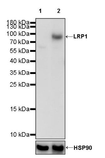
WB result of LRP1 Rabbit pAb
Primary antibody: LRP1 Rabbit pAb at 1/1000 dilution
Lane 1: MCF7 whole cell lysate 20 µg
Lane 2: A549 whole cell lysate 20 µg
Negative control: MCF7 whole cell lysate
Secondary antibody: Goat Anti-rabbit IgG, (H+L), HRP conjugated at 1/10000 dilution
Predicted MW: 85 kDa
Observed MW: 90 kDa
WB result of LRP1 Rabbit pAb
Primary antibody: LRP1 Rabbit pAb at 1/1000 dilution
Lane 1: C2C12 whole cell lysate 20 µg
Lane 2: mouse brain lysate 20 µg
Secondary antibody: Goat Anti-rabbit IgG, (H+L), HRP conjugated at 1/10000 dilution
Predicted MW: 85 kDa
Observed MW: 90 kDa
WB result of LRP1 Rabbit pAb
Primary antibody: LRP1 Rabbit pAb at 1/1000 dilution
Lane 1: C6 whole cell lysate 20 µg
Secondary antibody: Goat Anti-rabbit IgG, (H+L), HRP conjugated at 1/10000 dilution
Predicted MW: 85 kDa
Observed MW: 90 kDa
LRP1 Rabbit pAb at 1/50 dilution (1 µg) immunoprecipitating LRP1 in 0.4 mg U-87 MG whole cell lysate.
Western blot was performed on the immunoprecipitate using LRP1 Rabbit pAb at 1/1000 dilution.
Secondary antibody (HRP) for IP was used at 1/1000 dilution.
Lane 1: U-87 MG whole cell lysate 20 µg (Input)
Lane 2: LRP1 Rabbit pAb IP in U-87 MG whole cell lysate
Lane 3: Rabbit monoclonal IgG IP in U-87 MG whole cell lysate
Predicted MW: 85 kDa
Observed MW: 90 kDa
IHC shows positive staining in paraffin-embedded human cerebral cortex. Anti-LRP1 antibody was used at 1/200 dilution, followed by a HRP Polymer for Mouse & Rabbit IgG (ready to use). Counterstained with hematoxylin. Heat mediated antigen retrieval with Tris/EDTA buffer pH9.0 was performed before commencing with IHC staining protocol.
IHC shows positive staining in paraffin-embedded human placenta. Anti-LRP1 antibody was used at 1/200 dilution, followed by a HRP Polymer for Mouse & Rabbit IgG (ready to use). Counterstained with hematoxylin. Heat mediated antigen retrieval with Tris/EDTA buffer pH9.0 was performed before commencing with IHC staining protocol.
IHC shows positive staining in paraffin-embedded human tonsil. Anti-LRP1 antibody was used at 1/200 dilution, followed by a HRP Polymer for Mouse & Rabbit IgG (ready to use). Counterstained with hematoxylin. Heat mediated antigen retrieval with Tris/EDTA buffer pH9.0 was performed before commencing with IHC staining protocol.
IHC shows positive staining in paraffin-embedded mouse cerebral cortex. Anti-LRP1 antibody was used at 1/200 dilution, followed by a HRP Polymer for Mouse & Rabbit IgG (ready to use). Counterstained with hematoxylin. Heat mediated antigen retrieval with Tris/EDTA buffer pH9.0 was performed before commencing with IHC staining protocol.
IHC shows positive staining in paraffin-embedded rat cerebral cortex. Anti-LRP1 antibody was used at 1/200 dilution, followed by a HRP Polymer for Mouse & Rabbit IgG (ready to use). Counterstained with hematoxylin. Heat mediated antigen retrieval with Tris/EDTA buffer pH9.0 was performed before commencing with IHC staining protocol.
 您现在的位置: