
12 months from date of receipt / reconstitution, -20 °C as supplied
| 应用 | 稀释度 |
|---|---|
| IHC-P | 1:2000 |
| ICC | 1:500 |
CXCR2, or C-X-C chemokine receptor type 2, is a G protein-coupled receptor that binds to various chemokines, including CXCL1, CXCL2, CXCL3, CXCL5, CXCL6, CXCL7, and CXCL8 (also known as IL-8). It plays a significant role in the recruitment and activation of neutrophils during inflammation and is implicated in the pathogenesis of various diseases, including cancer. In the context of oncology, CXCR2 expression has been associated with tumor growth, angiogenesis, metastasis, and immune evasion. The receptor's interaction with its ligands can activate multiple signaling pathways, leading to the promotion of tumor-associated inflammation and immune suppression. Targeting CXCR2 and its signaling axis is being explored as a therapeutic strategy in cancer treatment, with potential applications in sensitizing tumors to immunotherapies and inhibiting cancer progression. Structural insights into CXCR2, such as those gained from cryo-electron microscopy and X-ray crystallography, are aiding in the development of small molecule inhibitors and other therapeutic agents.
IHC shows positive staining in paraffin-embedded human tonsil. Anti-CXCR2 antibody was used at 1/2000 dilution, followed by a HRP Polymer for Mouse & Rabbit IgG (ready to use). Counterstained with hematoxylin. Heat mediated antigen retrieval with Tris/EDTA buffer pH9.0 was performed before commencing with IHC staining protocol.
IHC shows positive staining in paraffin-embedded human liver. Anti-CXCR2 antibody was used at 1/2000 dilution, followed by a HRP Polymer for Mouse & Rabbit IgG (ready to use). Counterstained with hematoxylin. Heat mediated antigen retrieval with Tris/EDTA buffer pH9.0 was performed before commencing with IHC staining protocol.
IHC shows positive staining in paraffin-embedded human spleen. Anti-CXCR2 antibody was used at 1/2000 dilution, followed by a HRP Polymer for Mouse & Rabbit IgG (ready to use). Counterstained with hematoxylin. Heat mediated antigen retrieval with Tris/EDTA buffer pH9.0 was performed before commencing with IHC staining protocol.
IHC shows positive staining in paraffin-embedded human stomach. Anti-CXCR2 antibody was used at 1/2000 dilution, followed by a HRP Polymer for Mouse & Rabbit IgG (ready to use). Counterstained with hematoxylin. Heat mediated antigen retrieval with Tris/EDTA buffer pH9.0 was performed before commencing with IHC staining protocol.
IHC shows positive staining in paraffin-embedded human hepatocellular carcinoma. Anti-CXCR2 antibody was used at 1/2000 dilution, followed by a HRP Polymer for Mouse & Rabbit IgG (ready to use). Counterstained with hematoxylin. Heat mediated antigen retrieval with Tris/EDTA buffer pH9.0 was performed before commencing with IHC staining protocol.
IHC shows positive staining in paraffin-embedded human colon cancer. Anti-CXCR2 antibody was used at 1/2000 dilution, followed by a HRP Polymer for Mouse & Rabbit IgG (ready to use). Counterstained with hematoxylin. Heat mediated antigen retrieval with Tris/EDTA buffer pH9.0 was performed before commencing with IHC staining protocol.
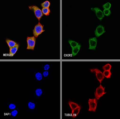
ICC shows positive staining in HCT116 cells. Anti-CXCR2 antibody was used at 1/500 dilution (Green) and incubated overnight at 4°C. Goat polyclonal Antibody to Rabbit IgG - H&L (Alexa Fluor® 488) was used as secondary antibody at 1/1000 dilution. The cells were fixed with 4% PFA and permeabilized with 0.1% PBS-Triton X-100. Nuclei were counterstained with DAPI (Blue). Counterstain with tubulin (Red).
 您现在的位置: