
12 months from date of receipt / reconstitution, -20 °C as supplied
| 应用 | 稀释度 |
|---|---|
| WB | 1:1000 |
| IHC-P | 1:250 |
CD6 is a transmembrane glycoprotein primarily found on the surface of T cells, serving as a co-stimulatory molecule that plays a significant role in T cell activation, proliferation, and differentiation. It binds to its ligands, CD166 (ALCAM) and CD318, which are expressed on various cell types, including antigen-presenting cells, epithelial, and endothelial tissues. The interaction of CD6 with its ligands modulates immune responses and is implicated in the pathogenesis of several autoimmune diseases. CD6 has been identified as a susceptibility locus in multiple sclerosis and Behcet’s disease, and its blockade using monoclonal antibodies, such as itolizumab, has shown clinical benefits in conditions like psoriasis. The therapeutic potential of targeting the CD6 pathway is currently being explored in various immune-mediated diseases, highlighting its importance in modulating T cell functions and maintaining immune homeostasis.
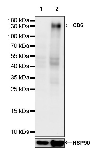
WB result of CD6 Recombinant Rabbit mAb
Primary antibody: CD6 Recombinant Rabbit mAb at 1/1000 dilution
Lane 1: HeLa whole cell lysate 20 µg
Lane 2: HuT 78 whole cell lysate 20 µg
Negative control: HeLa whole cell lysate
Secondary antibody: Goat Anti-rabbit IgG, (H+L), HRP conjugated at 1/10000 dilution
Predicted MW: 72 kDa
Observed MW: 140 kDa
This blot was developed with high sensitivity substrate
IHC shows positive staining in paraffin-embedded human tonsil. Anti-CD6 antibody was used at 1/250 dilution, followed by a HRP Polymer for Mouse & Rabbit IgG (ready to use). Counterstained with hematoxylin. Heat mediated antigen retrieval with Tris/EDTA buffer pH9.0 was performed before commencing with IHC staining protocol.
IHC shows positive staining in paraffin-embedded human spleen. Anti-CD6 antibody was used at 1/250 dilution, followed by a HRP Polymer for Mouse & Rabbit IgG (ready to use). Counterstained with hematoxylin. Heat mediated antigen retrieval with Tris/EDTA buffer pH9.0 was performed before commencing with IHC staining protocol.
Negative control: IHC shows negative staining in paraffin-embedded human cerebral cortex. Anti-CD6 antibody was used at 1/250 dilution, followed by a HRP Polymer for Mouse & Rabbit IgG (ready to use). Counterstained with hematoxylin. Heat mediated antigen retrieval with Tris/EDTA buffer pH9.0 was performed before commencing with IHC staining protocol.
IHC shows positive staining in paraffin-embedded Hodgkin’s lymphoma. Anti-CD6 antibody was used at 1/250 dilution, followed by a HRP Polymer for Mouse & Rabbit IgG (ready to use). Counterstained with hematoxylin. Heat mediated antigen retrieval with Tris/EDTA buffer pH9.0 was performed before commencing with IHC staining protocol.
IHC shows positive staining in paraffin-embedded human cervical squamous cell carcinoma. Anti-CD6 antibody was used at 1/250 dilution, followed by a HRP Polymer for Mouse & Rabbit IgG (ready to use). Counterstained with hematoxylin. Heat mediated antigen retrieval with Tris/EDTA buffer pH9.0 was performed before commencing with IHC staining protocol.
IHC shows positive staining in paraffin-embedded human lung squamous cell carcinoma. Anti-CD6 antibody was used at 1/250 dilution, followed by a HRP Polymer for Mouse & Rabbit IgG (ready to use). Counterstained with hematoxylin. Heat mediated antigen retrieval with Tris/EDTA buffer pH9.0 was performed before commencing with IHC staining protocol.
 您现在的位置: